Clin Endosc > Volume 46(2); 2013 > Article
Kwon and Ko: Balloon Catheter Assisted Biliary Stent Insertion
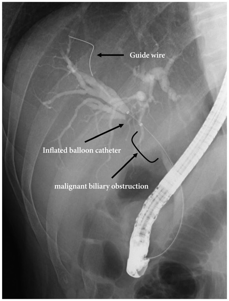
Endoscopic biliary stenting is widely used to palliate malignant obstruction or to treat benign biliary diseases. The technique is less invasive and guarantees high clinical success rates with low morbidity.1,2 In particular, a precise measurement of the length of stenotic area from the papilla makes it possible to place the stent successfully under fluoroscopic monitoring. Although previously taken abdominal computed tomography or magnetic resonance cholangiopancreatography might be helpful, simultaneous measurement using dedicated instruments during endoscopic retrograde cholangiopancreatography can be clinically most helpful.3 For this purpose, we employed a balloon catheter marked with a scale that allowed us to make direct endoscopic measurement of the length from the stenotic area to the papilla. The balloon catheter (Escort II double lumen extraction balloon; Cook Endoscopy, Winston-Salem, NC, USA), marked in advance with a scale, was one that we generally used as a retrieval catheter for biliary stone extraction. The followings are the step-by-step methods for measuring the length from the stenotic area to the papilla and stent insertion: 1) After the biliary stenosis was identified with cholangiography, a guide wire was inserted in advance through the stenosis; 2) The balloon catheter could pass through the stenosis over the guide wire and advance far enough along to pass the proximal margin of the stenosis; 3) Once the inflated balloon catheter was pulled back until it reached the proximal end of the stenosis (Fig. 1), we could endoscopically measure the length from the stenotic area to the papilla with the scale marked on the balloon cathether (Fig. 2); 4) After removing the balloon catheter, leaving the guide wire, a stent of adequate size was positioned through the stenosis.

NOTES

The authors have no financial conflicts of interest.

References

1. Speer AG, Cotton PB, Russell RC, et al. Randomised trial of endoscopic versus percutaneous stent insertion in malignant obstructive jaundice. Lancet 1987;2:57–62. 2439854.
crossref pmid
2. Cowling MG, Adam AN. Internal stenting in malignant biliary obstruction. World J Surg 2001;25:355–359. 11343193.
crossref pmid
3. Dumonceau JM, Heresbach D, Devière J, et al. Biliary stents: models and methods for endoscopic stenting. Endoscopy 2011;43:617–626. 21614754.
crossref pmid
Fig. 1
Cholangiography showing an obstruction of 3 cm in length at the proximal extrahepatic biliary tract. The first step begins with the insertion of a guide wire that is introduced inside the balloon catheter into the stenotic area. Once it has passed the stenotic area, the inflated balloon over the guide wire is withdrawn until it reaches the proximal margin of the stenosis.
ce-46-201-g001.jpg
Fig. 2
Endoscopic image of balloon catheter assisted measurement. The length from the stenotic area to the papilla is directly and precisely measured with the scale on the surface of the catheter. Inset; balloon catheter with marked scale. The balloon catheter was normally used as a retrieval catheter for biliary stone extraction and it was marked with the scale in advance.
ce-46-201-g002.jpg
TOOLS
PDF Links  PDF Links
PubReader  PubReader
ePub Link  ePub Link
XML Download  XML Download
Full text via DOI  Full text via DOI
Download Citation  Download Citation
  Print
Share:      
METRICS
1
Crossref
1
Scopus
4,086
View
35
Download
Related article
Delayed Severe Hemobilia after Endoscopic Biliary Plastic Stent Insertion   2016 May;49(3)
Editorial Office
Korean Society of Gastrointestinal Endoscopy
#817, 156 Yanghwa-ro (LG Palace, Donggyo-dong), Mapo-gu, Seoul, 04050, Korea
TEL: +82-2-335-1552   FAX: +82-2-335-2690    E-mail: CE@gie.or.kr
Copyright © Korean Society of Gastrointestinal Endoscopy.                 Developed in M2PI
Close layer