INTRODUCTION
Crohn disease (CD) is a chronic inflammatory disease that mainly affects the gastrointestinal (GI) tract. It may also have various extraintestinal manifestations such as uveitis, arthritis, pyoderma gangrenosum, erythema nodosum, episcleritis, sclerosing cholangitis, and hemolytic anemia [1-3]. Symptomatic tracheobronchial involvement is not common, although CD with chronic bronchitis [4], bronchiectasis [4,5], upper airway obstruction [6,7], granulomatous interstitial pneumonitis, bronchiolitis obliterans [4], granulomatous lung disease [4,8,9], and interstitial lung disease [10] has been reported. This report describes a case of CD with tracheal involvement.
CASE REPORT
An 18-year-old Korean woman was diagnosed with CD 4 months ago. She received prednisolone at 50 mg/day for 1 month; after tapering and discontinuation of prednisolone, mesalazine at 3 g/day was administered for maintenance. She visited a primary clinic because of prolonged purulent coughing, fever, and dyspnea. She had never smoked and had no history of environmental or occupational exposure to agents that might cause respiratory distress. At the time of her visit, the Montreal classification of CD was A2L3B1, and the CD activity index was 71.92. She received antibiotics for 5 days along with symptomatic treatment, but her dyspnea and fever persisted, and stridor was found upon physical examination. She was then referred to our hospital for further evaluation.
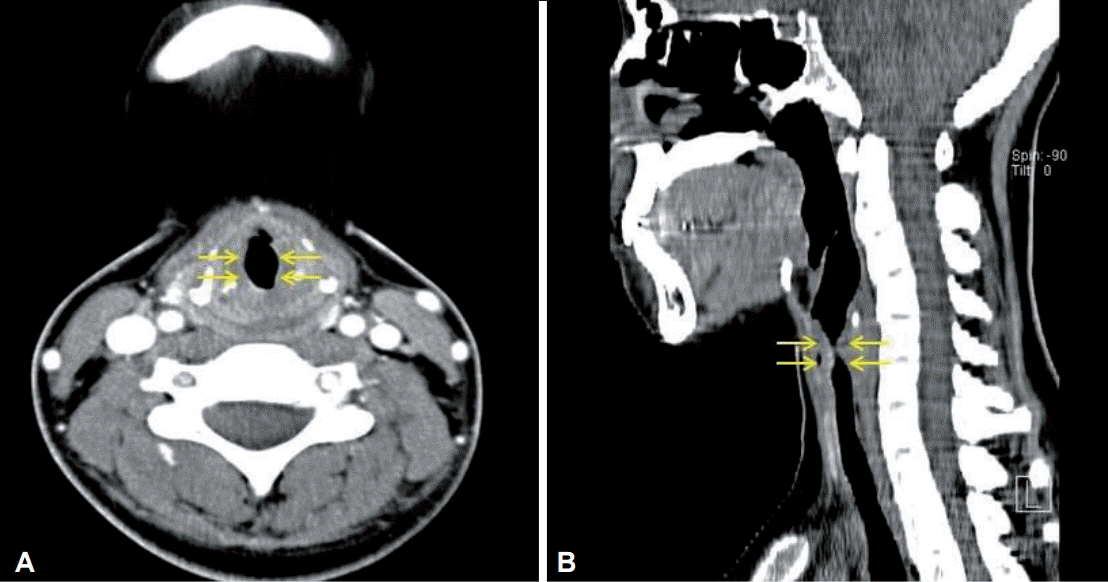
A physical examination revealed stridor in her neck. Her blood pressure was 110/70 mm Hg, her heart rate was 100 beats per minute, her body temperature was 36.7o C, and her peripheral capillary oxygen saturation level was 97% (room air). The electrocardiogram and chest radiographs were normal. Laboratory data on admission included the following: white blood cells, 18,770/خ¼L (polymorphonuclear neutrophils, 67.6%; eosinophils 0.4%); hemoglobin, 10.4 g/dL; platelets, 628,000/خ¼L; and C-reactive protein, 5.75 mg/dL. The remaining serum chemistry results and the coagulation profile were normal. Computed tomography of the neck performed to determine the cause of the stridor and cough revealed circumferential wall thickening of the larynx and hypopharynx (Fig. 1).
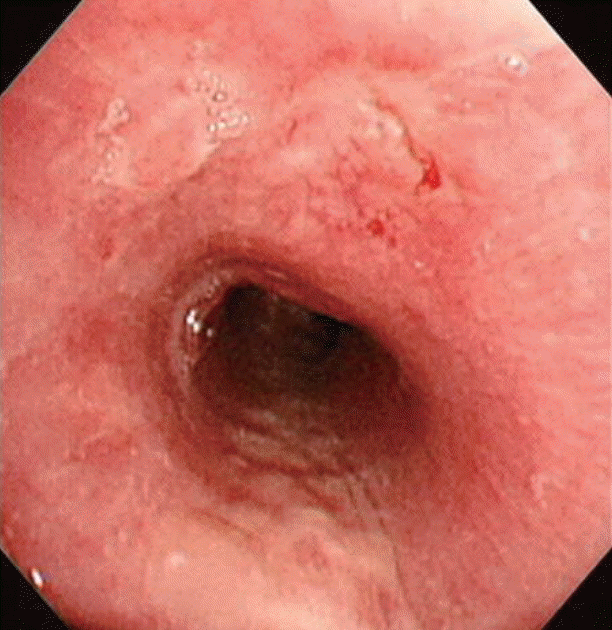
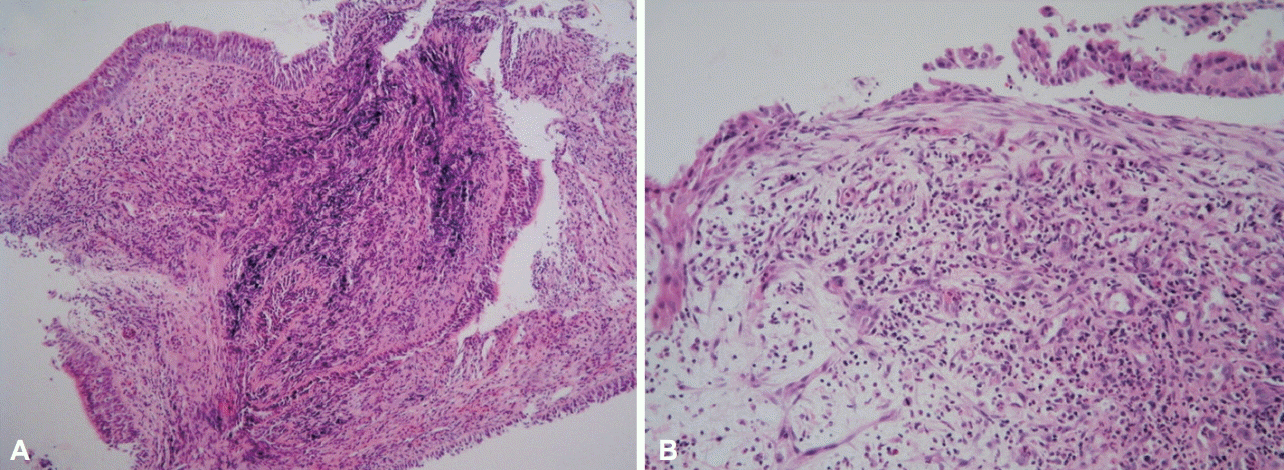
We initially thought that the patient had bacterial tracheitis and therefore administered antibiotics (amoxicillin/clavulanate, 3.6 g/day) for 4 days. However, her symptoms and stridor did not improve. Flexible fibrotic bronchoscopy (FFB) showed mucosal irregularity, edema, and a yellowish patch-like mucosal lesion on the proximal part of trachea (Fig. 2). The results of bacterial culturing, acid-fast bacilli staining of the culture, and tuberculosis polymerase chain reaction (TB-PCR) assays of the bronchial washing fluid were negative, as were those of interferon-gamma release assays (IGRAs) for Mycobacterium tuberculosis. A bronchoscopic biopsy showed non-specific inflammatory reactions including dense inflammatory cell infiltration, necrotic detritus due to tissue destruction, and granulation tissue indicating healing of damaged tissue. However, the biopsy specimen contained no granulomas, which are relatively well known histopathologic features of CD (Fig. 3). Owing to the negative results of the tests, we suspected CD with tracheal involvement and therefore began administering intravenous methylprednisolone at 1 mg/kg per day.
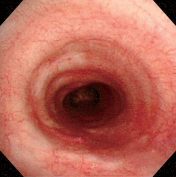
Four days later, her cough and dyspnea were relieved, and the fever and stridor also disappeared. After 2 months, bronchoscopy revealed marked improvement in the tracheal lesion (Fig. 4). The patient was treated with an oral corticosteroid for 4 weeks, and more than 1 year later, there has been no recurrence of the CD-associated lung involvement.
DISCUSSION
We describe the first case in Korea of tracheobronchial involvement in CD, which is rare even globally. Since Kraft et al. [5] reported bronchopulmonary involvement in inflammatory bowel disease in 1976, only 13 cases including the present case (seven men and six women, ages 18 to 59 years; average age, 28 years) [3] have been reported, and the patient in our case is the youngest (Table 1).
In the present case, diseases that could cause similar clinical symptoms (e.g., bacterial tracheitis, tracheal tuberculosis, and sarcoidosis) as tracheobronchial involvement were excluded. Bacterial tracheitis was eliminated owing to negative results in the bacteriologic studies and unresponsiveness to an antibacterial agent. Mycobacterial infection was excluded for the following reasons: no growth of mycobacteria in repeated samplings of sputum and the bronchial washing fluid, negative results in an IGRA and nested TB-PCR, and the absence of caseating granulomas. Sarcoidosis was ruled out because there was no evidence of extrapulmonary sarcoid involvement or typical sarcoid granulomas in bronchial biopsies.
Tracheobronchial involvement of CD is rare, although several cases have been reported [1,2,5,6,8,9,11-13]. Similar to previous reports [2,6,9,13,14] and to the colonoscopic findings for CD, the bronchoscopic findings in our case were mucosal irregularity, elevation, edema, and the presence of a yellowish patch-like lesion. In the present case, as in previous cases, the pathologic findings of the bronchial biopsy were epithelial and subepithelial infiltrates of diverse types of inflammatory cells including neutrophils, macrophages, lymphocytes, and plasma cells [2,6,9,13,14]. According to Asami et al. [3], bronchoscopic and pathologic findings such as these indicate that tracheobronchial inflammation results from the activation of macrophages and T lymphocytes, as does colonic inflammation. Previous reports also suggest that a common antigen triggers mucosal immune responses in both the GI tract and airway [2,6,9,13,14]. In our case, when the tracheal involvement in CD was exacerbated, the colonic condition of the patient was stabilized. Thus, the clinical courses of tracheobronchial involvement and GI involvement appear to be dissimilar [3,11]. This may be because the inflammatory response is amplified differently in the respiratory tract and GI tract, even though it may be triggered by the same antigen in both locations [3,11]. Patients are usually diagnosed with CD before presentation of pulmonary manifestations; however, airway involvement has been reported in patients without a diagnosis of CD [15]. Pathologically, airway infiltration by inflammatory cells and mucosal ulceration may occur. However, non-caseating granulomas are not always present, as in cases of colon involvement in CD [4,6]. In our case, there was only granulation tissue, not granulomas.
It has been reported that both inhaled and systemic corticosteroids effectively treat upper airway involvement in CD. Six previous cases were treated with oral steroids [2,5,8,12], two with inhaled steroids [3,9] and four with oral and inhaled steroids (Table 1) [1,6,11,13]. Both respiratory and GI symptoms were improved in all patients receiving steroids regardless of the method of administration [3]. In our case, we used intravenous steroids, because the patient refused oral steroids owing to nausea, abdominal pain, and a sore throat; she also had problems using inhaled corticosteroids. We began administering intravenous methylprednisolone at 1 mg/kg per day 4 days after detecting tracheal involvement; her symptoms improved thereafter, and after 2 months, the FFB findings were markedly improved.
The clinical course of tracheobronchial involvement in CD tends to be more responsive to corticosteroids than does lower respiratory tract involvement such as bronchiectasis in CD [4]. This is especially true when high dose systemic corticosteroids are administered early in the course of upper respiratory tract involvement [16]. Clinical sequelae of residual tracheal stenosis and respiratory failure have been reported with delayed diagnosis and consequent delayed treatment with systemic steroids [2,6,12].
We report the first Korean case of tracheobronchial involvement in CD and show a dramatic response to systemic steroids. Our case suggests that pulmonary involvement should be considered in CD patients with unexplained respiratory symptoms.


