INTRODUCTION
Endoscopic variceal ligation (EVL) is the preferred endoscopic treatment method for esophageal variceal bleeding and is associated with increased survival and minimized rebleeding and complication rates [1,2]. Potential complications include chest pain, bleeding, stricture formation, aspiration pneumonia, dysphagia, and perforation. The incidence of these complications, however, is low [3].
CASE REPORT
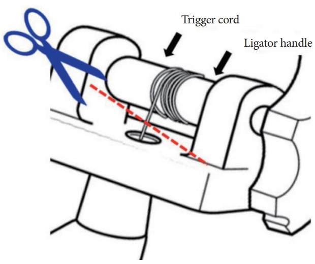
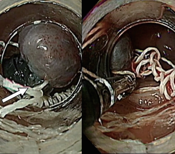
A 46-year-old man with liver cirrhosis due to chronic hepatitis B was admitted to our center after an episode of melena occurring 7 hours prior. At the time of admission, his vital signs were normal and stable. He had a history of EVL for esophageal variceal bleeding 1 year previously. The results of the complete blood count and serum biochemical test were as follows: white blood cells, 4,700/╬╝L; hemoglobin, 9.9 g/dL; hematocrit, 31.0%; platelets, 118,000/╬╝L; prothrombin time, 1.47 international normalized ratio; activated partial thromboplastin time, 40.7 sec; aspartate aminotransferase, 24 IU/L; alanine aminotransferase, 21 IU/L; albumin, 3.6 g/dL; and total bilirubin, 2.4 mg/dL. Endoscopic band ligation (6 Shooter Saeed Multi-Band Ligator; Cook Medical, Bloomington, IN, USA) was performed without sedation. During additional band ligation, the trigger cord malfunctioned and was inserted between the bands on a single varix (Supplementary Video 1). The trigger cord was stuck between several bands, and the endoscope could not be retrieved. Forcibly moving the endoscope would have meant tearing the varix and potentially causing massive bleeding. Thus, we had to think of another way to solve the problem immediately. After cutting the trigger cord with scissors (it was connected to the ligator handle) (Fig. 1), we pushed the cord out of the endoscope inside the accessory channel using forceps. After selectively grasping the trigger cord between the bands (Fig. 2), we gradually advanced the endoscope and forceps to pull out the trigger cord between the bands (Fig. 3). After the trigger cord was removed from between the bands, the endoscope was released and retrieved. One month after the procedure, the patient did not experience any hemorrhagic events and received an additional EVL procedure.
DISCUSSION
Our case has been reviewed and investigated by the device manufacturer. They collected all the other ligators that were in stock at that time and experimented to determine if the same event would occur again. However, all the other products worked normally.
In conclusion, the definitive cause of the incident could not be determined through the evaluation of the recalled products or provided video. We set up the device appropriately. The trigger cord on the device appeared to be in the correct position between each band, and the deployment beads were positioned correctly on the device. Therefore, we concluded that this was a rare event that occurred by chance. To our knowledge, this is the first reported case wherein a malfunctioning multiple-band ligator could have led to a potentially serious damage to the esophageal varix, with the risk of massive bleeding. We safely removed the equipment using scissors and forceps.