Clin Endosc > Volume 54(3); 2021 > Article
Sasaki, Mie, and Sasahira: Blind Spot of Enteral Stenting for Malignant Duodenal Obstruction due to Colon Cancer: A Rare Case of Duodenocolonic Fistula
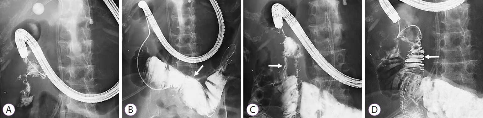
A 57-year-old woman had undergone right hemicolectomy, partial pancreatic head resection, and partial duodenectomy for advanced ascending colon cancer. Owing to a local recurrence, she was referred to our hospital. Percutaneous transhepatic biliary drainage was performed for a malignant biliary obstruction, and the local recurrence was treated with radiotherapy. Systemic chemotherapy was then started, but the patient had a complication of malignant duodenal obstruction 1 year after the recurrence. Only non-contrast-enhanced abdominal computed tomography could be obtained because of the patient’s allergy to the contrast medium (Fig. 1). The size of the recurrence site was estimated to be approximately 5 cm. However, detailed information on the occlusion site was difficult to obtain before the endoscopic treatment. A forward-viewing endoscope was inserted in the duodenum, and the guidewire was advanced through the stenosis (Fig. 2A). Subsequent contrast imaging revealed a dilated intestinal tract (Fig. 2B). However, contrast imaging at the stenosis revealed another lumen, and the first lumen was clarified to be the colon, and the guidewire was passed through the duodenocolonic fistula (Fig. 2C). The guidewire was turned to enter another lumen, a true duodenal lumen (Fig. 2D). As the enteral stent was placed at the papilla of Vater, an uncovered enteral stent (NEXENT Duodenal Pyloric Stent 22 mm × 12 cm; Next Biomedical, Incheon, Korea) was placed at the duodenal obstruction. After the procedure, the patient could take a normal diet (Fig. 3A, B).
Duodenocolonic fistula is a rare complication. It is sometimes caused by benign diseases such as chronic inflammation, a foreign body, and iatrogenic diseases (e.g., those induced by radiotherapy) [1-3]. Duodenocolonic fistula due to malignancy is rare [4]. Furthermore, malignant duodenal obstruction due to colon cancer is unusual. When inserting an enteral stent, the stenosis is passed through using the guidewire under fluoroscopy. The guidewire is confirmed to be inside the intestinal tract based on the contrast image and course of the guidewire. In the case of a duodenocolonic fistula, the obstruction usually occurs at the infraduodenal angle. As the guidewire needs to be advanced to the third portion of duodenum, its course is quite similar to that of the transverse colon. Misplacement of an enteral stent is one of the most critical complications. To avoid this complication, clinicians should be aware that the distal bowel tract below the stenosis is usually collapsed and that no residue-like material is visible. Understanding that the guidewire may go through a lumen different from the assumed duodenum when the fistula is formed is also necessary. The present case of a duodenocolonic fistula was a blind spot in the placement of a duodenal stent. Information on such a case is useful for clinicians who perform enteral stenting.

NOTES

Conflicts of Interest: The authors have no potential conflicts of interest.
Funding: None.
Author Contributions
Data curation: Takashi Sasaki, Takafumi Mie, Naoki Sasahira
Writing-original draft: TS
Writing-review&editing: TM, NS

Fig. 1.
Non-contrast-enhanced abdominal computed tomography image showing a local recurrence at the duodenum, which caused a malignant gastric outlet obstruction. The arrow indicates the local recurrence of the colon cancer.
ce-2020-140f1.jpg
Fig. 2.
(A) An endoscope is inserted in the duodenum, and the guidewire is advanced through the stenosis. (B) The contrast image shows a dilated intestinal tract. The arrow indicates the lumen of the transverse colon. (C) The contrast image of the stenosis shows another lumen. The first lumen is confirmed to be a duodenocolonic fistula, which is indicated by the arrow. (D) The guidewire is then advanced to another lumen, and the contrast image shows the duodenum. The lumen indicated by the arrow is the true duodenal lumen.
ce-2020-140f2.jpg
Fig. 3.
An uncovered enteral stent at the duodenal obstruction. (A) Abdominal radiograph. (B) Endoscopic view.
ce-2020-140f3.jpg

REFERENCES

1. Liu S, de Blacam C, Lim FY, Mattei P, Mamula P. Magnetic foreign body ingestions leading to duodenocolonic fistula. J Pediatr Gastroenterol Nutr 2005;41:670–672.
crossref pmid
2. Park MS, Kim WJ, Huh JH, et al. Crohn’s duodeno-colonic fistula preoperatively closed using a detachable endoloop and hemoclips: a case report. Korean J Gastroenterol 2013;61:97–102.
crossref pmid
3. Javia SB, Patel R, Singhal S. Endoscopic closure with an over-the-scope clip of a duodenocolonic fistula caused by a migrated biliary stent. Gastrointest Endosc 2016;83:845–846; discussion 846-847.
crossref
4. Okuda KV, Laass M, Schuchardt K, et al. Duodenocolonic fistula as a rare complication of intestinal Burkitt lymphoma in a three-year-old boy. Klin Padiatr 2018;230:138–141.
crossref pmid pdf
TOOLS
PDF Links  PDF Links
PubReader  PubReader
ePub Link  ePub Link
XML Download  XML Download
Full text via DOI  Full text via DOI
Download Citation  Download Citation
  Print
Share:      
METRICS
0
Crossref
0
Scopus
2,898
View
66
Download
Related articles
Endoscopic Stenting for Malignant Biliary Obstruction: Results of a Nationwide Experience  2021 September;54(5)
Insertion of Self Expandable Metal Stent for Malignant Stomal Obstruction in a Patient with Advanced Colon Cancer  2012 November;45(4)
Editorial Office
Korean Society of Gastrointestinal Endoscopy
#817, 156 Yanghwa-ro (LG Palace, Donggyo-dong), Mapo-gu, Seoul, 04050, Korea
TEL: +82-2-335-1552   FAX: +82-2-335-2690    E-mail: CE@gie.or.kr
Copyright © Korean Society of Gastrointestinal Endoscopy.                 Developed in M2PI
Close layer