Clin Endosc > Volume 54(6); 2021 > Article
Kim, Lee, Lee, and Park: Endoscopic Submucosal Dissection of an Inverted Pyloric Gland Adenoma Using Dental Floss and Clip Traction
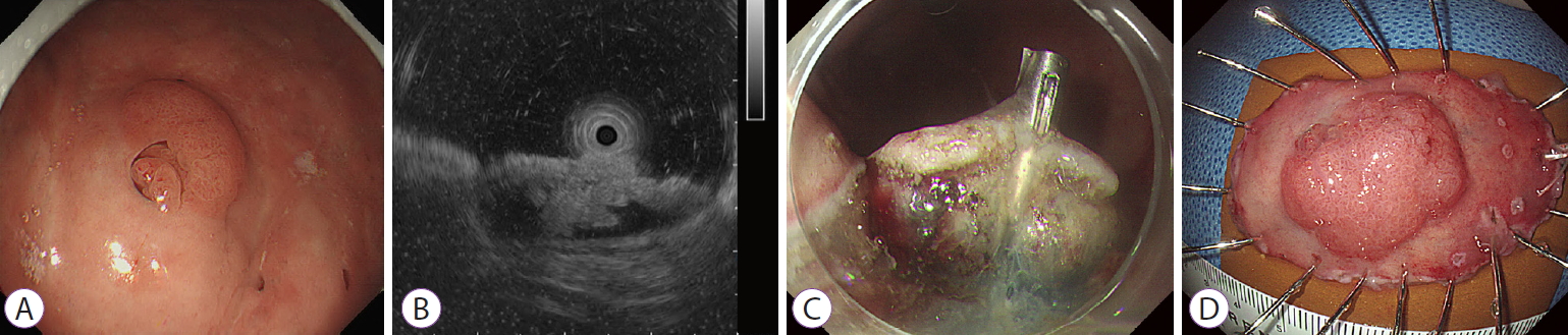
A gastric adenoma with low-grade dysplasia was incidentally detected during screening endoscopy in a 52-year-old woman. The endoscopy revealed a reddish elevated lesion with a diameter of 1.5 cm and an opening on the greater curvature of the upper body (Fig. 1A). Endoscopic ultrasonography of the lesion revealed a downward papillary growth into an anechoic cystic space of approximately 2 cm in size in the deep mucosal and submucosal layers (Fig. 1B). Endoscopic submucosal dissection (ESD) was performed for complete removal of the adenoma and accompanying cystic lesion (Supplementary Video 1).
After the borders of the tumor were marked, a 0.9% saline solution with indigo carmine and epinephrine was injected in the submucosal layer. Circumferential mucosal incision and submucosal dissection were then performed using a dual knife and an insulated-tipped knife. During dissection, a clip attached with dental floss was used to secure the dissection plane between the tumor and the proper muscle layer (Fig. 1C). The tumor was then completely resected (Fig. 1D).
Histopathological examination revealed that the cystic invagination of the mucosal lesion was composed of neoplastic glands with high-grade dysplasia. Most of the glandular cells had round nuclei with prominent nucleoli and mucinous columnar cytoplasm that resemble the pyloric glandular epithelium (Fig. 2). Therefore, the lesion was diagnosed as an inverted pyloric gland adenoma with high-grade dysplasia.
Pyloric gland adenoma is a rare neoplasm, and the histological resemblance of the tumor to the deep glands of the gastric mucosa near the pylorus is reflected in its name [1]. It occurs more frequently in women and in the elderly. As adenocarcinoma is detected in approximately 30% of pyloric gland adenomas [2], they are considered precancerous lesions, and endoscopic removal is indicated [3]. As described in our report, ESD using dental floss and clip traction seems to be an efficient and safe technique for inverted pyloric gland adenomas, especially when the tumor is located in the greater curvature of the upper or middle stomach [4].

NOTES

Conflicts of Interest
The authors have no potential conflicts of interest.
Funding
None.

Supplementary Material

Video 1. Endoscopic submucosal dissection of an inverted pyloric gland adenoma (https://doi.org/10.5946/ce.2020.164.v001).

Fig. 1.
(A) A reddish elevated lesion with a diameter of 1.5 cm and an opening on the greater curvature of the gastric upper body. (B) Endoscopic ultrasonography of the lesion showing a downward papillary growth into an anechoic cystic space of approximately 2 cm in size in the deep mucosal and submucosal layers. (C) During endoscopic submucosal dissection, the dissection plane is secured between the tumor and the proper muscle layer using a clip attached with dental floss. (D) The resected specimen.
ce-2020-164f1.jpg
Fig. 2.
(A) Histopathological examination showing that the cystic invagination of the mucosal lesion is composed of neoplastic glands with high-grade dysplasia (hematoxylin and eosin stain ×12.5). (B) Most of the glandular cells have round nuclei with prominent nucleoli and mucinous columnar cytoplasm that resemble the pyloric glandular epithelium (hematoxylin and eosin stain ×200).
ce-2020-164f2.jpg

REFERENCES

1. Golger D, Probst A, Wagner T, Messmann H. Pyloric-gland adenoma of the stomach: case report of a rare tumor successfully treated with endoscopic submucosal dissection. Endoscopy 2008;40(Suppl 2):E110–E111.
crossref pmid pdf
2. Oberhuber G, Stolte M. Gastric polyps: an update of their pathology and biological significance. Virchows Arch 2000;437:581–590.
crossref pmid pdf
3. Yamamoto M, Nishida T, Nakamatsu D, Adachi S, Inada M. Endoscopic findings of inverted pyloric gland adenoma resected by endoscopic submucosal dissection. J Gastrointestin Liver Dis 2018;27:361.
crossref pmid
4. Yoshida M, Takizawa K, Suzuki S, et al. Conventional versus traction-assisted endoscopic submucosal dissection for gastric neoplasms: a multicenter, randomized controlled trial (with video). Gastrointest Endosc 2018;87:1231–1240.
crossref pmid
TOOLS
PDF Links  PDF Links
PubReader  PubReader
ePub Link  ePub Link
XML Download  XML Download
Full text via DOI  Full text via DOI
Download Citation  Download Citation
Supplement  Supplement
  Print
Share:      
METRICS
2
Web of Science
3
Crossref
2
Scopus
4,268
View
157
Download
Related articles
Waterjet-assisted endoscopic submucosal dissection in the colorectum: safe and effective?  2022 November;55(6)
Efficacy and Safety of Endoscopic Submucosal Dissection for Superficial Gastric Neoplasms: A Latin American Cohort Study  2022 March;55(2)
Long-Term Outcomes of Endoscopic Submucosal Dissection of Undifferentiated-Type Early Gastric Cancer  2021 March;54(2)
Endoscopic Submucosal Dissection versus Surgery for Undifferentiated-Type Early Gastric Cancer: A Systematic Review and Meta-Analysis  2021 March;54(2)
Endoscopic Submucosal Dissection of a Colonic Calcifying Fibrous Tumor  2020 July;53(4)
Editorial Office
Korean Society of Gastrointestinal Endoscopy
#817, 156 Yanghwa-ro (LG Palace, Donggyo-dong), Mapo-gu, Seoul, 04050, Korea
TEL: +82-2-335-1552   FAX: +82-2-335-2690    E-mail: CE@gie.or.kr
Copyright © Korean Society of Gastrointestinal Endoscopy.                 Developed in M2PI
Close layer