INTRODUCTION
Diagnosing unclear lymphadenopathy such as lymph node metastasis has an enormous impact on the treatment of neoplastic diseases. Therefore, the aim of treatment usually switches from cure to palliation. Numerous other diseases are accompanied by lymphadenopathy, including benign conditions such as infections and inflammatory disease [1]. Hence, sampling of the target node is often required to unveil the underlying diagnosis and to enable adequate clinical management [2].
Intrathoracic or intraabdominal lymphadenopathies are clinically challenging because the nodes are difficult to sample using non-invasive techniques. Therefore, invasive methods such as mediastinoscopy or laparotomy are needed but are associated with risks [3]. A mini-invasive method such as endosonography (EUS) is an alternative approach in such scenarios [4]. Endosonographic characteristics of malignant lymph nodes include large size, hypoechogenicity, distinct border, round shape, and high tissue stiffness on elastography [5,6]. Nevertheless, the lymph node appearance on EUS alone is not sufficient to firmly distinguish benign nodes from malignant ones [7]. Instead, pathology is required to confirm the underlying etiology.
Very few prospective studies have addressed the utility of EUS-guided tissue sampling in lymphadenopathies. The diagnostic accuracy of EUS-guided fine-needle aspiration (EUS-FNA) is generally moderate [8,9] but poor in diagnosing lymphomas [10]. EUS-guided fine-needle biopsy sampling (EUS-FNB) can provide histological specimens and is a sensitive and safe method to use in solid pancreatic and subepithelial lesions [11,12]. However, the role of EUS-FNB in the diagnosis of lymphadenopathy remains unclear and there is a lack of high-quality, prospective studies on this topic.
Therefore, the aim of this study was to evaluate the diagnostic accuracy of EUS-FNB in relation to EUS-FNA in the workup of intrathoracic and intraabdominal lymphadenopathies of unclear etiology. An additional aim was to investigate whether target-node or sampling-related factors had any impact on the outcome of sampling.
MATERIALS AND METHODS
Study setting and design
This study was conducted at the endoscopy unit of a tertiary care center, Sahlgrenska University Hospital (GEA-SUH), between January 2014 and October 2019. In this prospective study, we consecutively enrolled patients aged Ōēź18 years who were referred for EUS-guided sampling. All subjects initially presented with enlarged intrathoracic or intraabdominal lymph nodes (Ōēź20 mm) of unclear etiology, as visualized on cross-sectional imaging when other modalities for sampling were considered non-feasible. Patients were excluded from the study if they refused to participate or if no diagnostic EUS-guided sampling was performed.
The study was approved by the Regional Ethical Review Board of Gothenburg (Dnr: 1092-11), and the study protocol conforms to the ethical guidelines of the 1975 Declaration of Helsinki (6th Revision, 2008). Written informed consent was obtained from all included patients. The study was registered at ClinicalTrials.gov (NCT02360839). The STARD protocol was applied throughout the study.
The EUS-procedure
The study subjects were examined by one of two experienced endosonographers (PH and Riadh Sadik). A linear echoendoscope (EG3870UTK; Pentax, Tokyo, Japan) and an ultrasound processor (HI VISION Ascendus; Hitachi, Tokyo, Japan) were used to examine the patients under deep sedation. Before the start of the procedure, the patients were randomized to a first pass with FNA or FNB. We anticipated that the first pass might cause damage to the lymph node tissue and impair the yield of the second pass. Therefore, we aimed to exclude the possibility that the diagnostic accuracy of either technique (FNA or FNB) was better if performed as a first-pass rather than a second-pass modality.
After the identification of the target node, relevant features were recorded, including lesion size, shape, vascularization, and echogenicity. Parameters related to the EUS procedure were also recorded. Then, the endosonographer performed dual sampling with EUS-FNA and EUS-FNB in the same lymph node. The first needle pass was performed according to the results of the randomization and the second procedure was performed with the other needle.
Sampling with FNA for cytological analysis was performed using a 25-gauge open-tip needle (EchoTip®; Wilson-Cook Medical, Limerick, Ireland or Expect Slimline®; Boston Scientific, Marlborough, MA, USA), while sampling with FNB for histopathological analysis was performed with a 22-gauge reverse-beveled needle (Procore®; Wilson-Cook Medical). In both EUS-FNA and EUS-FNB, standard suction and sampling by fanning were performed [13]. Any technical failures or instant adverse events, such as bleeding, were recorded.
If available, a cytotechnician was present on-site to prepare and preliminarily assess the yield of EUS-FNA. The first part of the first aspirate was smeared onto glass slides, airdried, and stained with Giemsa for rapid on-site cytology evaluation (ROSE). All residual FNA materials were put in a methanol-based solution (ThinPrep®; Hologic, Marlborough, MA, USA) for the preparation of cell blocks. Additional FNA passes were performed if the initial aspirate was inadequate. No samples were put into saline, and flow cytometry was not performed in any case.
The yield of the EUS-FNB was put directly into formalin tubes and the FNB core was assessed visually by the endosonographer. If the initial FNB core was considered too sparse upon gross examination, one or two additional FNB passes were performed. There was no fixed number of passes for either needle. Relevant clinical information and information on the target lesion characteristics on EUS were supplied to the pathology department.
Cytopathology and histopathology
The FNA material stored in ThinPrep┬« was used for the preparation of cell blocks using an automatized system (CellientTM; Hologic, Marlborough, MA, USA) according to the manufacturerŌĆÖs instructions. The cell blocks were paraffin-embedded, cut into thin sections, and placed on slides. Prior to immunostaining, the slides were fixed in acetone at ŌĆÆ20┬░C for 10 min, then treated in a microwave oven (at 700 W for 7 min and 300 W for 15 min) with a Dako Target Retrieval Solution (citrate pH 6, S2031), cooled to room temperature, and rinsed with deionized water.
The FNB core biopsy samples were formalin-fixed and paraffin-embedded. Sections (3ŌĆÆ4 ┬Ąm) were placed on positively charged glass slides and treated in Dako PT-Link using EnVisionTM FLEX Target Retrieval Solution (TRS High).
If necessary, the samples were further analyzed by immunocytochemistry and immunohistochemistry, respectively. No fixed panel of antibodies was used, but instead, the primary monoclonal antibodies were selected based on the preliminary results of Giemsa or hematoxylin-eosin staining and the suspected diagnosis in each case. Immunostaining was performed using a Dako Autostainer Link using EnVisionTM FLEX according to the manufacturerŌĆÖs instructions (DakoCytomation). Positive and negative controls were included in each run.
After the sample processing, the quality of the FNA and FNB samples was assessed by a pathologist trained in the assessment of samples acquired via EUS. The specimens were classified as adequate (specimens with sufficient material for morphology assessment and any immunohistochemistry analysis) or as inadequate (specimens with few target node cells or with contaminating material). Finally, each adequate sample was given a specific diagnosis and was categorized as benign (negative for malignancy), malignant (positive for malignancy), highly suggestive for malignancy, or non-diagnostic [14].
Samples were regarded as diagnostic for malignancy if the pathology report was positive for malignancy or highly suggestive of malignancy. In cases with a final diagnosis of lymphoma or sarcoidosis, the samples were assessed by a hematopathologist (VC) blinded to the final diagnosis. The WHO 2016 Classification of tumors of hematopoietic and lymphoid tissues was applied to assess and classify samples suspicious of lymphomas [15]. Given that in most cases, lymphoma can be conclusively confirmed only by obtaining and processing an entire node, samples were categorized by the study pathologist as either suggestive of lymphoma or non-diagnostic for lymphoma. Sarcoidosis was diagnosed based on the presence of non-necrotizing granulomas.
In cases with a benign final diagnosis, a sample was regarded as diagnostic only if the quality of the sample was adequate, that is, a sample containing benign or reactive cells/tissue from the target node. If the sample was acellular or contained entirely contaminating cells, it was regarded as non-diagnostic, that is, it was not used to calculate the diagnostic specificity.
Follow-up, final diagnosis, and reference standard
All patients were monitored after EUS via clinical follow-up by the referring clinician and by the study investigators until the final diagnosis was established (minimum 6 months) or until death (follow-up <6 months). The follow-up included a minimum of one visit to the outpatient unit of the referring department and a minimum of one computed tomography/magnetic resonance imaging examination (except for cases sent directly for surgery). Potential adverse events were checked at the follow-up visit. In addition, the study investigators screened the complete medical files of all study patients at 30 days post-EUS.
Specifically, any adverse event related to EUS (<30 days post-EUS), the imaging results, and the results of any diagnostic procedure post-EUS were recorded. Adverse events were categorized according to the validated ClavienŌĆōDindo classification [16].
The final diagnosis of each patient was determined based on the reference standard, which was primarily the pathology of the surgical specimens, pathology of any other diagnostic sampling, or pathology of the autopsy. In patients not subjected to any of these procedures, the clinical diagnosis at a minimum of six months of follow-up was used as the reference standard.
Study outcomes
The primary outcome was the diagnostic sensitivity, specificity, and overall accuracy of EUS-FNB and EUS-FNA, respectively. The secondary outcome was the adverse event rate of EUS-guided sampling.
Statistical analysis
Descriptive continuous data were described as medians and interquartile ranges (IQR), while descriptive categorical data were described as frequencies.
In dual sampling procedures, McNemarŌĆÖs test for paired data with a binary outcome was used to test for differences comparing EUS-FNB vs. EUS-FNA and EUS-FNA/B (both techniques combined) vs. EUS-FNA. In the final calculation of the diagnostic sensitivity, specificity, and accuracy, an intention-to-diagnose analysis was performed, that is, all cases were counted, including cases with no yield. FisherŌĆÖs exact test was used to analyze any factor with a potential impact on the primary outcome.
Based on local, unpublished data on the accuracy of EUS-FNA, a sample size calculation was performed (statistical power: 80%, alpha error: 0.05) to detect a 20% superiority in the diagnostic accuracy of EUS-FNB compared with that of EUS-FNA (paired proportions), indicating 45 patients were required.
A two-tailed p-value <0.05 was regarded as statistically significant for all analyses. All the statistical calculations and tests were performed using SPSS Statistics version 25.0 (IBM Co., Armonk, NY, USA).
RESULTS
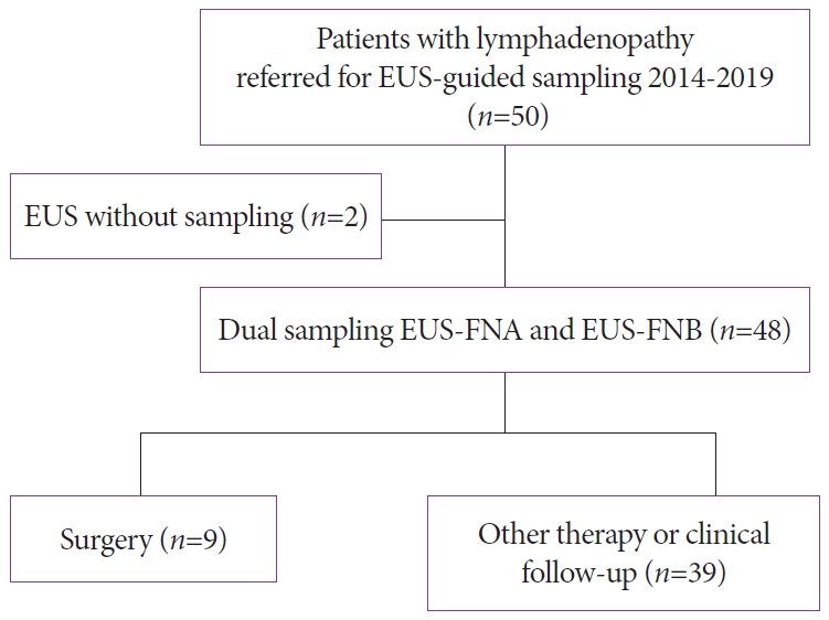
A total of 50 consecutive eligible patients with lymphadenopathy were referred to GEA-SUH. Finally, EUS-guided dual sampling EUS-FNA and EUS-FNB were performed in 48 patients (median age: 69 years [IQR, 59ŌĆō76]; 24/48 females [50%]). Two patients did not undergo sampling due to a highly vascularized target region with a high risk of bleeding in one case and due to lack of any detected lymphadenopathy on EUS in the other case (Fig. 1). The study baseline characteristics are presented in Table 1. The median clinical follow-up time was 16 months (IQR, 6ŌĆō24 months) with a minimum follow-up of six months in all patients with a benign final diagnosis.
The primary study outcome
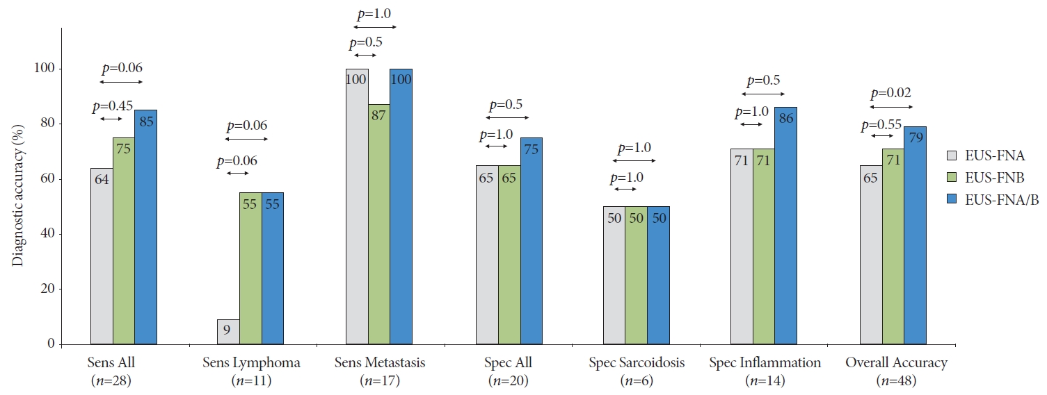
The diagnostic accuracy of EUS-FNA and EUS-FNB were comparable, and both modalities were highly sensitive for metastatic nodes (Fig. 2). EUS-FNB was borderline significantly more sensitive for lymphoma (Fig. 2).
The combination of both techniques (EUS-FNA/B) was more accurate and more sensitive than EUS-FNA alone with borderline significance (Fig. 2).
A non-significant higher number of needle passes was recorded in EUS-FNA than in EUS-FNB (2.40 [95% confidence interval, 2.17ŌĆō2.63] vs. 2.02 [95% confidence interval, 1.73ŌĆō2.31]; p=0.066). The order of sampling did not affect the accuracy of EUS-FNA (FNA first: 16/24 [67%] vs. FNB first: 14/24 [58%], p=0.76) or the accuracy of EUS-FNB (FNA first: 16/24 [67%] vs. FNB first: 18/24 [75%], p=0.75).
No factors were recorded with a significant impact on the diagnostic accuracy of EUS-FNA or EUS-FNB (Table 2).
DISCUSSION
This work is the first prospective study comparing the performance of EUS-FNA and EUS-FNB specific to lymphadenopathy. Both modalities had a high sensitivity for metastatic nodes, while EUS-FNB showed a tendency to be more sensitive in cases of lymphoma. Both approaches were safe, and no adverse events were recorded.
Others have investigated the diagnostic capacity of EUS-FNB performed with various types of needles. However, with few exceptions, there is a remarkable lack of high-quality, high-volume studies on patients with lymphadenopathy [17]. In a systematic review published in 2016 [18], the authors analyzed studies comparing EUS-FNB performed with a reverse-beveled biopsy needle and routine EUS-FNA. Out of nine identified studies, only three studies were enrolled, comprising a small number of patients with lymphadenopathy [18-21]. Consequently, few conclusions on the diagnostic capacity of EUS-FNB in lymphadenopathy or comparisons to EUS-FNA could be drawn from this meta-analysis.
In a recent large multicenter study including 608 patients, the diagnostic accuracy of EUS-FNB was found to be significantly superior to that of EUS-FNA (82% vs. 72%) [22]. Various target lesions were included, with lymphadenopathy constituting a minority. Only 13 patients (2%) had a final diagnosis of lymphoma. No details on the sensitivity of EUS-FNB in this specific subgroup of patients were provided. The criteria for a diagnostic sample were determined by the principal investigator at each participating site, and no information was provided on the criteria applied for the diagnosis of lymphoma. Moreover, the FNB needle used in the aforementioned study was a 20 G forward-bevel needle, making the results somewhat challenging to compare with those of our study [22].
In 2020, de Moura et al. published a retrospective study on EUS-FNB in patients with lymphadenopathy exclusively [23]. A total of 209 patients were included in the study and subsequently subjected to EUS-FNB (n=101) or EUS-FNA (n=108). Needles of various brands and sizes were used, and no predefined study protocol was applied. The overall diagnostic accuracy and sensitivity of EUS-FNB and EUS-FNA were 83% and 75% and 79% and 67%, respectively. Importantly, the findings by de Moura et al. are also somewhat difficult to compare with other studies since the distribution of the final diagnoses was not presented [23]. Moreover, the criteria applied to the diagnostic sample were not provided.
In numerous malignancies, the detection and microscopic confirmation of a metastatic lymph node are crucial to determine the clinical management of the patient. For example, liver transplantation should not be performed if malignant nodes are detected in a patient with cholangiocarcinoma. As mentioned above, only a few prospective studies focusing on EUS-FNB have addressed the issue of lymph node metastases. Meanwhile, several retrospective studies have analyzed the accuracy of EUS-FNA in lymphadenopathy [24]. Results from some prospective studies show that EUS-FNA is indeed sensitive for metastatic nodes [25], which was confirmed in the current study. We posit that EUS-FNA is an adequate sampling technique when metastasis is suspected with no apparent need for a switch to EUS-FNB.
According to the results of our study, the utility of the 22 G reverse-beveled EUS-FNB needles seems lower in lymphoma than in metastatic nodes. However, in 6 out of 11 cases with lymphoma, histology specimens from EUS-FNB were indeed highly suspicious for lymphoma (diffuse large B-cell lymphoma). In 5 out of these 6 cases, the hematologist decided to start chemotherapy without additional diagnostic procedures because the target nodes were difficult to access for alternative approaches. On the other hand, the EUS-FNA performed poorly and was non-diagnostic for lymphoma in all but one case. In 2012, Gimeno-Garc├Ła et al. published a review of the available literature investigating EUS-FNA in suspected lymphoma [17]. Few studies were identified, and only one study, including 6 patients, was prospective. Yasuda et al. reported high sensitivity (97%) in the diagnosis of lymphoma when using a large 19 G EUS-FNA needle [26]. However, this result was achieved by combining cytopathology, flow cytometry, and cytogenetic analysis. The sensitivity of cytopathology was reported to be as low as 52% [26]. Thus, the use of EUS-FNA in the diagnosis of lymphoma can be questioned [15,27,28].
Indeed, the combination of both techniques, EUS-FNA and EUS-FNB, was statistically more accurate than EUS-FNA alone in the present study. However, in metastatic nodes, EUS-FNB added no diagnostic information compared with EUS-FNA alone; in addition, in lymphoma, EUS-FNA added no diagnostic information compared with EUS-FNB alone. Therefore, in most scenarios, the use of both techniques in lymphadenopathy seems to have no utility or value, and such an approach would only increase costs and procedure time.
The number of needle passes required to obtain a diagnostic yield is often presented as a variable in the literature. Hypothetically, a low number of passes may lead to a shorter procedural time and a lower adverse event rate. In the current study, we reported a lower mean number of needle passes in EUS-FNB compared to that in EUS-FNA with borderline significance. This finding is in line with previous studies on EUS-FNB [21,23,29,30].
ROSE is costly, and there is no clear consensus on the necessity of ROSE in EUS-guided sampling [23,31-34]. The results of the present study show no incremental diagnostic effect of ROSE in EUS-FNA, which may indicate that EUS-FNB is an attractive alternative to EUS-FNA in lymphadenopathies even in centers benefitting from access to ROSE. On the other hand, EUS-FNB has the obvious drawback that FNB cores are not well aimed for on-site smears and preliminary microscopic assessment of samples.
Regarding patient safety during EUS-FNB and dual sampling procedures in lymphadenopathy, we did not record any adverse events in any of the study patients within 30 days post-EUS. This finding is novel and serves endosonographers with valuable information. Previously, reverse-beveled EUS-FNB needle sampling has been found safe for subepithelial and pancreatic lesions [12,35]. Apparently, EUS-FNB can be regarded as safe as EUS-FNA in the sampling of lymphadenopathy [17].
In the interpretation of results from a diagnostic study, it is important to keep in mind the criteria applied to categorize diagnostic samples [14,36,37]. Unfortunately, there are discrepancies among publications on EUS-guided sampling. As in the study by van Riet et al. [22], we decided to accept and categorize samples highly suspected for malignancy as diagnostic for malignancy. Others, like Han et al. [38], have applied stricter criteria and have categorized samples assessed as suspicious for malignancy as non-diagnostic in the calculation of diagnostic sensitivity. Meanwhile, in the current study, we applied a strict intention-to-diagnose approach and required that samples from benign nodes should contain not only benign material but also benign cells from the target node (lymphocytes). A sample containing cells from the trajectory line only, such as the gastrointestinal epithelium, was considered non-diagnostic. Such criteria will automatically decrease the diagnostic specificity and consequently the overall diagnostic accuracy in an intention-to-diagnose analysis. A per-protocol analysis would have resulted in a higher diagnostic accuracy.
To the best of our knowledge, the current study is the largest prospective study on EUS-FNB to exclusively include patients with lymphadenopathy. Moreover, valuable and specific data on the diagnostic sensitivity for metastases and lymphoma are presented. The study is also strengthened by the single-center setting, which limits the risk of assignment bias and heterogeneity of the included cases. A hematologist and hematopathologist were involved in the accurate assessment of the specimens and their clinical implications. Finally, the long clinical follow-up accounts for high reliability concerning the final diagnosis of the study cases.
Some limitations of the current study should be taken into consideration. For practical reasons, the pathologists could not be blinded to the sampling modality performed. Only a minority of the study cases had a final diagnosis based on pathology. However, this is to be expected in all lymphadenopathy cohorts since neither a majority of patients with benign nodes nor a majority of patients with metastatic disease should be referred for surgery. In addition, we meticulously screened all medical files of the study participants during the follow-up period. Nevertheless, the lack of comparative pathology in some of the study cases should be taken into consideration when interpreting the results of the current study. Further, there exist FNB needles with an alternative design, such as the Franseen-tip needle and the fork-tip needle, both with proven high utility in a variety of lesions [39,40]. However, when designing the current study, there were fewer needles available and our ambition was to evaluate one specific needle and sampling technique since the use of several different FNB-needles would have made the results more difficult to interpret.
In conclusion, EUS-guided fine-needle biopsy sampling performed with a reverse-beveled needle is safe in lymphadenopathy of unknown etiology. The diagnostic accuracies of EUS-FNB and EUS-FNA are comparable, and both techniques are highly sensitive for metastatic nodes. The diagnosis of lymphoma may be facilitated by the use of EUS-FNB instead of EUS-FNA, but this remains challenging for endosonographers, regardless of the needle used.