Clin Endosc > Volume 45(2); 2012 > Article
Kim, Ryu, Kim, Lee, Im, Huh, Choi, and Kang: Cecal Fecaloma Due to Intestinal Tuberculosis: Endoscopic Treatment

Abstract

Colorectal fecaloma is a mass of accumulated feces that is much harder in consistency than a fecal impactation. The rectosigmoid area is the common site for fecalomas and the cecum is the most unusual site. Diagnosis is usually made by distinctive radiographic findings of a mobile intraluminal mass with a smooth outline and no mucosal attachment. Most of the fecalomas are successfully treated by conservative methods such as laxatives, enemas and rectal evacuation. When conservative treatments have failed, endoscopic procedures or a surgical intervention may be needed. We report here that a cecal fecaloma caused by intestinal tuberculosis scar was successfully removed by endoscopic procedures.

INTRODUCTION

Fecaloma is a laminated mass of accumulated feces that is much harder in consistency than a fecal impaction.1 It is usually located in the sigmoid colon or rectum, but rarely in the cecum.2-4 Diagnosis is usually made from radiographic findings of a mobile intraluminal mass with a smooth outline and no mucosal attachment.2,5 Most fecalomas are successfully treated by conservative methods such as laxatives, enemas and rectal evacuation.2 When conservative treatments have failed, a surgical intervention may be needed.6 Only one case treated by an endoscopic procedure has recently been reported.7 We report here a case of cecal fecaloma, associated with an intestinal tuberculosis scar, that was successfully removed by endoscopic balloon dilatation of the stricture and mechanical destruction of the fecaloma with a polypectomy snare and grasping forceps.

CASE REPORT

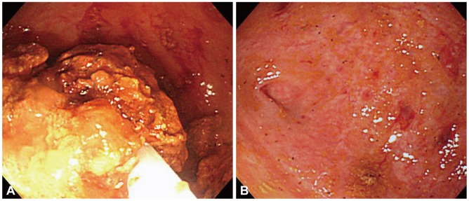
A 30-year-old female presented with a 2-month history of intermittent pain and a palpable mass in the right lower quadrant of the abdomen. She had a history of chronic constipation with about 2 bowel movements per week and hard stools. Five years earlier, she had undergone an appendectomy. She was diagnosed with pulmonary tuberculosis 9 months ago and was taking antituberculosis therapy. Her height was 163 cm and her weight was 49 kg. Physical examination revealed mild abdominal tenderness and a ping-pong ball-sized movable mass in the right lower quadrant of the abdomen. An abdominal computed tomography (CT) scan showed a 3.0-cm, round, laminated intraluminal mass with calcification in the cecum (Fig. 1A, B). Colonoscopy revealed fibrotic scar tissue probably due to intestinal tuberculosis in the ascending colon and a web-like stricture in the cecum around a patulous ileocecal valve. In a blind space which was formed by the stricture, a yellowish mass was found (Fig. 2). We could not insert a fiberoptic colonoscope into the cecum, and dilatation was thus performed using a th-rough-the-scope balloon (CRE balloon; Boston Scientific Co., Marlborough, MA, USA) with a diameter of 12 to 15 mm on inflation (Fig. 3). After endoscopic balloon dilatation, the colonoscope was able to pass into the cecum, and a 3.0-cm, yellowish fecaloma was observed. We broke down the fecaloma with a polypectomy snare and grasping forceps. The fecaloma was successfully removed by using a water jet and grasping forceps through the endoscopic procedure (Fig. 4A, B; Supplementary Video 1 online). There was no ulcer in the cecal base. Three mo-nths later, her symptoms improved, and there was no evidence of fecaloma recurrence.

DISCUSSION

Although fecal impaction is a common condition, fecaloma is an extremely rare form of impaction that refers to an accumulation of fecal material which forms a mass separable from the rest of the bowel contents.1 Fecaloma is found most frequently in the rectum or sigmoid because stools in the left colon become firmer and colon diameter is smaller on the left side than on the right side.6 The cecum is an unusual site, and only 4 cases have been reported in the English literature.2-4
There are several causes of fecaloma, and they have been described in patients suffering with chronic constipation, Hi-rschsprung's disease, Chagas' disease, and psychiatric diseases.8-10 It is thought that our case of fecaloma developed due to chronic constipation and prolonged impaction of fecal material in a pouch which was formed by stricture. This is the first case of fecaloma that was associated with an intestinal tuberculosis scar.
Diagnosis of fecaloma is usually made radiologically from a characteristic intraluminal mass seen on plain X-rays, barium enema and abdominal CT.2,5 The mass has smooth margins, some mobility within the bowel lumen and no attachment to the mucosal surface.
Complications of fecaloma are obstruction, ulceration, bleeding and perforation of the colon as well as hydronephrosis.11 Treatments include laxatives, enemas, rectal evacuation, surgical intervention and endoscopic removal.6 This is the second case of fecaloma that was removed successfully by the endoscopic procedure and the first case of cecal fecaloma that was removed successfully by the endoscopic procedure with endoscopic balloon dilatation.

NOTES

The authors have no financial conflicts of interest.

References

1. Garisto JD, Campillo L, Edwards E, Harbour M, Ermocilla R. Giant fecaloma in a 12-year-old-boy: a case report. Cases J 2009;2:127. 19196473.
crossref pmid pmc
2. Cid AA, Pietruk T, Bidari CZ, Ehrinpreis MN. Cecal fecaloma mimicking colonic neoplasm. Dig Dis Sci 1981;26:1134–1137. 7307862.
crossref pmid
3. Gilbert RF. Cecal infarction secondary to a distal obstructing fecaloma: association with drug abuse. South Med J 1980;73:1296–1297. 6106293.
crossref pmid
4. Lasser A, Conte M, Solitare GB. Stercoraceous perforation of the cecum: report of two cases. Dis Colon Rectum 1975;18:410–412. 1157644.
crossref pmid
5. Kantarci M, Fil F. Education and imaging. Gastrointestinal: fecaloma in a dilated sigmoid colon. J Gastroenterol Hepatol 2007;22:955. 17565653.
crossref pmid
6. Sakai E, Inokuchi Y, Inamori M, et al. Rectal fecaloma: successful treatment using endoscopic removal. Digestion 2007;75:198. 17893439.
crossref pmid
7. Freud WI, Zikmund A, Stroud CS, Fries JW. Fecaloma: report of a case and review of the literature. Gastroenterology 1955;29:446–452. 13262500.
crossref pmid
8. Kim KH, Kim YS, Seo GS, Choi CS, Choi SC. A case of fecaloma resulting in the rectosigmoid megacolon. Korean J Neurogastroenterol Motil 2007;13:81–85.

9. Campbell JB, Robinson AE. Hirschsprung's disease presenting as calcified fecaloma. Pediatr Radiol 1973;1:161–163. 4773697.
crossref pmid
10. Araki T, Miki C, Yoshiyama S, Toiyama Y, Sakamoto N, Kusunoki M. Total proctocolectomy and ileal J-pouch anal anastomosis for chagasic megacolon with fecaloma: report of a case. Surg Today 2006;36:277–279. 16493541.
crossref pmid
11. Knobel B, Rosman P, Gewurtz G. Bilateral hydronephrosis due to fecaloma in an elderly woman. J Clin Gastroenterol 2000;30:311–313. 10777195.
crossref pmid
Fig. 1
An abdominal computed tomography scan (A, axial view; B, coronal view) shows a 3.0-cm, round, laminated intraluminal mass with calcification in the cecum.
ce-45-174-g001.jpg
Fig. 2
Colonoscopy reveals a fibrotic scar and a web-like stricture in the cecum with a yellowish mass in a blind space which was formed by stricture.
ce-45-174-g002.jpg
Fig. 3
Endoscopic balloon dilatation with a through-the-scope balloon.
ce-45-174-g003.jpg
Fig. 4
(A) Colonoscopic view of breaking down the fecaloma with a polypectomy snare. (B) Colonoscopy reveals the lumen of the cecum after the fecaloma was successfully removed by the endoscopic procedure.
ce-45-174-g004.jpg
TOOLS
PDF Links  PDF Links
PubReader  PubReader
ePub Link  ePub Link
XML Download  XML Download
Full text via DOI  Full text via DOI
Download Citation  Download Citation
  Print
Share:      
METRICS
5
Crossref
4
Scopus
10,124
View
56
Download
Related articles
Clinical meaning of sarcopenia in patients undergoing endoscopic treatment  
Recent Developments in Devices Used for Gastrointestinal Endoscopy Sedation  2021 March;54(2)
Obesity and Endoscopic Treatment  2011 May;42(5)
Crohn's Disease vs. Intestinal Tuberculosis  2010 March;42(29)
A Case of Duodenal Fistula Caused by Intestinal Tuberculosis  2004 March;28(3)
Editorial Office
Korean Society of Gastrointestinal Endoscopy
#817, 156 Yanghwa-ro (LG Palace, Donggyo-dong), Mapo-gu, Seoul, 04050, Korea
TEL: +82-2-335-1552   FAX: +82-2-335-2690    E-mail: CE@gie.or.kr
Copyright © Korean Society of Gastrointestinal Endoscopy.                 Developed in M2PI
Close layer