Tribonias, Voudoukis, Vardas, Theodoropoulou, Margarita-Eleni, and Paspatis: Endoscopic Retrograde Cholangiopancreatography-Related Large Jejunal Perforation: Operate or Apply Over-the-Scope Clip Device?
To the Editor:
We have read with interest the review by Lee et al. 1 titled "Endoscopic treatments of endoscopic retrograde cholangiopancreatography-related duodenal perforations." Herein, we would like to describe the management of an analogous case from among our experience of 3,000 endoscopic retrograde cholangiopancreatographies (ERCPs) in 8 years. An 84-year-old man underwent ERCP because of recurrent cholangitis with intrahepatic bile duct dilatation, as seen on MRCP. The patient underwent a Billroth II gastrojejunostomy for the treatment of gastroduodenal ulcer disease 30 years earlier. During ERCP, multiple attempts to introduce the scope were made because of difficulty in approaching the papilla. An endoscope-related perforation was visualized at the end of the afferent loop ( Fig. 1). The diameter of the defect was approximately 20 mm, and we first contemplated nonoperative closure with an over-the-scope clip (OTSC) device. 1 Despite the existence of published data 2, 3 reporting successful endoscopic closure of large duodenal perforations by OTSC, we felt that it was necessary to perform a computed tomography (CT) scan to rule out the presence of any substantial intraperitoneal fluid collection. The abdominal CT was performed 30 minutes after the recognition of the perforation, and demonstrated pneumoperitoneum with fluid collection around the afferent loop ( Fig. 2). All fluids previously detected in the afferent loop had been carefully suctioned at the time of insertion, although type I duodenal perforations caused by the endoscope tend to be large with persistent fluid leaks in the retroperitoneal or intraperitoneal space. 4 Therefore, we decided that, despite the patient's advanced age, an operation would be preferable to the application of an OTSC device in this case. Laparotomy revealed a peritoneal cavity full of bilious fluid. The patient underwent a thorough washout of the abdominal cavity, surgical closure of the defect, and drainage. Recovery was uneventful and he was discharged 10 days later. On the basis of the findings of the present case, we would like to suggest that OTSC application could be considered an optimal treatment for duodenal perforations in inoperable patients or in patients who are not septic and have minimal peritoneal fluid collection. However, we believe that in cases involving a septic patient with intraperitoneal fluid collection, an endoscopic closure is probably unsuitable. Prompt surgical intervention with washout, closure of the perforation, and drainage is crucial for achieving recovery without sepsis or abscess formation.
Fig.┬Ā1
Large jejunal perforation in the afferent loop with direct visualization of the peritoneal cavity. 
Fig.┬Ā2
Computed tomography performed 1 hour after the perforation showing the presence of pneumoperitoneum with fluid collections around the afferent loop. 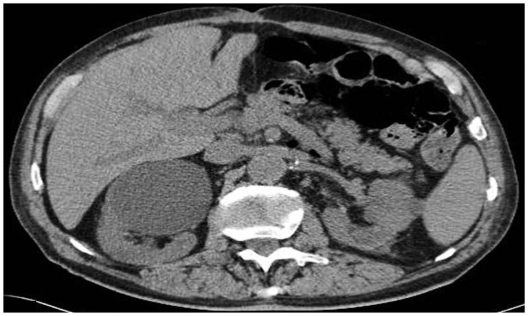
To the Editor:
Thank you for your interest in our research, and we agree with your opinion. As you know, the choice of treatment for endoscopic retrograde cholangiopancreatography (ERCP)-related gut perforation, which is not that simple, has a large impact on a patient's prognosis. Consequently, we need to be careful about choosing treatment methods for perforation. Before choosing surgical or endoscopic treatment, several factors should be considered, such as perforation size, risk of peritonitis, and status of the patient. In your presented case, the size of the jejunal perforation was large (about 20 mm) and might have been caused by diagnostic endoscopy itself, which usually causes a large perforation. The subsequent abdominal computed tomography (CT) scan for observing the amount of pneumoperitoneum and fluid collection in the peritoneal cavity showed lots of bile content influx to the peritoneal cavity. Moreover, the patient underwent Billroth II gastrojejunostomy. They diagnosed the perforation early during examination and then checked the CT scan; they had also been careful about suctioning during the insertion. According to the primary treatment principle, authors successfully performed early surgical closures. Therefore, we agree with your decision for surgical closure, which seems to have been a reasonable choice for that situation.
However, the patient was advanced in age (84-year-old), and he may have had risk factors for surgical treatment, aside from age, including a history of previous abdominal surgery. So, avoiding surgical treatment may have been the best choice. I think that the two important points for decision were the time difference between making and finding the perforation, and the degree of fluid leakage in the retroperitoneal or intraperitoneal space. Early recognition and management is essential for the prognosis. Recent studies have shown a successful fistula or perforation closure with an over-the-scope clip (OTSC) in clinical cases, 1, 2 and one study demonstrated successful closure with an OTSC of up to a 2-cm perforation in the stomach and a 3-cm perforation in the colon. 3 We are now researching closure methods, including the OTSC system, for large colon perforations. Based on our preliminary data, the average procedure time for closure of a large perforation with an OTSC is less than 5 minutes. This may be faster than what we expected. An OTSC as well as the endoloop technique combined with endoclips can be applied in larger perforations in selected cases. Although nonsurgical suturing therapies are not yet widely accepted as the primary treatment for ERCP-related perforation, our opinion is that you can also try an endoscopic treatment first, such as the OTSC or other endoscopic closure using an endoloop with endoclips, followed by a check with a CT scan. Then we can carefully check the physical examination, patient symptoms, and lab findings. After assessing these factors, if surgical treatment is needed, we should not hesitate to do surgery. As authors mentioned, if the patient is septic and there is a large amount of intraperitoneal fluid collection, early surgical management is essential. Endoscopic closure should be performed by experienced endoscopists as early as possible following recognition of perforation. However, endoscopic therapy cannot totally replace surgical closure. We should choose the best option based on the characteristics of the perforation, such as size and anatomical site, the patient's condition, and available expertise or circumferences.
References
1. Weiland T, Fehlker M, Gottwald T, Schurr MO. Performance of the OTSC System in the endoscopic closure of iatrogenic gastrointestinal perforations: a systematic review. Surg Endosc 2013;27:2258ŌĆō2274. 23340813.   2. Di┼¤ibeyaz S, K├Čksal A┼×, Parlak E, Torun S, ┼×a┼¤maz N. Endoscopic closure of gastrointestinal defects with an over-the-scope clip device. A case series and review of the literature. Clin Res Hepatol Gastroenterol 2012;36:614ŌĆō621. 22704818. 3. Matthes K, Jung Y, Kato M, Gromski MA, Chuttani R. Efficacy of full-thickness GI perforation closure with a novel over-the-scope clip application device: an animal study. Gastrointest Endosc 2011;74:1369ŌĆō1375. 21981814.
|
|


