Skip Navigation
Skip to contents

Clin Endosc : Clinical Endoscopy

OPEN ACCESS

Articles

Page Path
HOME > Clin Endosc > Volume 47(6); 2014 > Article
Case Report Successful Treatment of a Gastric Plasmacytoma Using a Combination of Endoscopic Submucosal Dissection and Oral Thalidomide
Se Young Park1, Hee Seok Moon1, Jae Kyu Seong1, Hyun Yong Jeong1, Beum Yong Yoon1, Se Woong Hwang1, Kyu Sang Song2
Clinical Endoscopy 2014;47(6):564-567.
DOI: https://doi.org/10.5946/ce.2014.47.6.564
Published online: November 30, 2014

1Department of Internal Medicine, Chungnam National University School of Medicine, Daejeon, Korea.

2Department of Pathology, Chungnam National University School of Medicine, Daejeon, Korea.

Correspondence: Hee Seok Moon. Department of Internal Medicine, Chungnam National University School of Medicine, 266 Munhwa-ro, Jung-gu, Daejeon 301-747, Korea. Tel: +82-42-280-7164, Fax: +82-42-254-4553, mhs1357@cnuh.co.kr
• Received: October 31, 2013   • Revised: December 28, 2013   • Accepted: January 13, 2014

Copyright © 2014 Korean Society of Gastrointestinal Endoscopy

This is an Open Access article distributed under the terms of the Creative Commons Attribution Non-Commercial License (http://creativecommons.org/licenses/by-nc/3.0/) which permits unrestricted non-commercial use, distribution, and reproduction in any medium, provided the original work is properly cited.

  • 6,132 Views
  • 51 Download
  • 7 Web of Science
  • 7 Crossref
prev next
  • We report a rare case of a gastric plasmacytoma treated with endoscopic resection and oral thalidomide therapy. A 70-year-old man was admitted to our hospital with indigestion. He had no specific medical history and unremarkable laboratory results. Gastroendoscopic findings revealed a focal, erythematous, flat elevated lesion in the anterior wall of the stomach antrum. A biopsy revealed atypical lymphocytes. Endoscopic submucosal dissection (ESD) with an insulation-tipped knife was performed 45 days after diagnosis. Radiological and hematological evaluations, including a bone marrow biopsy, were performed and showed no involvement of other organs. The patient was diagnosed with extramedullary gastric plasmacytoma. Follow-up gastroendoscopy was performed three times during a 2-year period and showed nonspecific ESD scarring. The patient's condition was found to be stable.
Extramedullary plasmacytomas (EMPs) are uncommon and occur almost exclusively in the head, neck, and upper respiratory tract; EMPs in gastrointestinal organs are rare.1 The next most frequent site of mass lesion occurrence is the stomach; however, this is also extremely rare, accounting for less than 5% of all EMPs.2 EMPs of the head and neck are more sensitive to radiotherapy than to surgery.2,3 Endoscopic submucosal dissection (ESD) is a less invasive therapeutic approach for early gastric cancer and has been accepted as a standard therapeutic approach for early gastric cancers limited to the mucosa.4 Here, we report the successful treatment via ESD of a patient with a solitary gastric EMP confined to the mucosa.
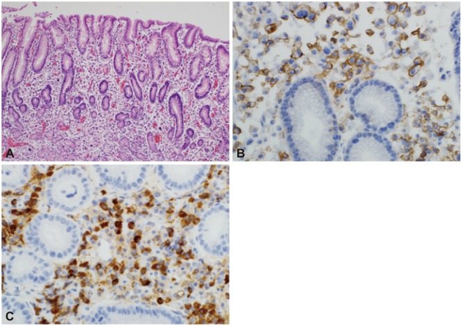
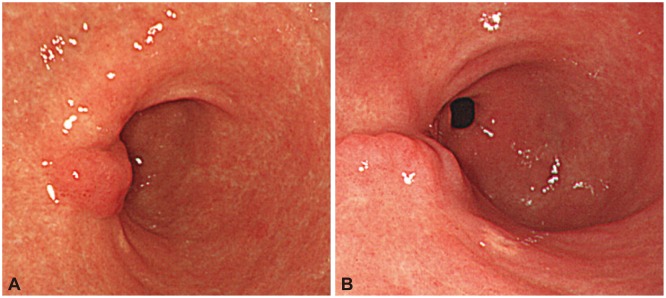
A 70-year-old man was admitted to our hospital with the complaint of dyspepsia. He had no previous specific medical history and unremarkable laboratory results. We performed gastroscopy and observed a flat lesion with focal erythematous changes in the anterior wall of the antrum (Fig. 1). There were no specific serum and urine immunoelectrophoresis or immunofixation findings. A biopsy revealed poorly differentiated neoplastic cells and atypical lymphocytes, consistent with metastatic carcinoma. A Giemsa stain indicated Helicobacter pylori negativity. To rule out cancer and systemic diseases such as lymphoma, we performed a bone marrow aspiration, biopsy, and peripheral blood smear. There was no evidence of clonal marrow or peripheral plasmacytosis. We also performed abdominal and pelvic computed tomography (CT) and positron emission tomography/CT scans; these yielded unremarkable results, particularly with regard to lymph node and bone lesions. Regarding the gastric focal lesion, we performed an endoscopic procedure to confirm the previous endoscopic biopsy result as well as for therapeutic reasons using an insulation-tipped knife (KD-610L; Olympus, Tokyo, Japan) (Fig. 2). After successful ESD, the acquired specimen revealed plasma cell infiltration into the lamina propria; however, these cells did not extend deeply into the submucosal layer. The lesion was confined to the mucosa and had a clear resection margin. Numerous plasma cells with atypical hyperchromatic nuclei were observed to infiltrate the gastric mucosa. The neoplastic cells were positive for CD138 (Fig. 3).
We finally diagnosed the patient with gastric EMP. Because EMPs are systemic, the patient was treated with additional oral thalidomide and dexamethasone. Later, we performed three endoscopic follow-up exams during a 2-year period and confirmed nonspecific ESD scarring. We found the patient's condition to be stable (Fig. 4).
Plasma cell neoplasms are classified into four types: solitary myeloma (bone plasmacytoma), multiple myeloma (bone marrow and other systemic involvements), extramedullary (soft tissue) plasmacytoma, and plasmablastic sarcoma.5 EMP is defined as an immunoproliferative, monoclonal, plasma cell tumor that develops in an extramedullary organ; gastric plasmacytomas are a very rare form of EMP. Gastric tumors account for 2% to 5% of all EMPs and tend to be identified at a late stage if an endoscopic examination is not performed.6 Gastric plasmacytomas can be classified into infiltrative, nodular, ulcerative, and polypoid types, of which the nodular type is most common. Most gastric plasmacytomas are large, deeply infiltrating tumors with ulceration; however, tumor cells are limited to the mucosal and submucosal layers in the early stage.7 Almost all patients with EMPs are treated with radiation therapy, surgery, or combination therapy (surgery and/or chemotherapy or irradiation). However, no general treatment guidelines have been established for gastric EMPs. Additionally, the invasiveness of these initial therapeutic approaches precludes their recommendation for EMPs of the head and neck.2,3
We also considered the possibility of mucosa-associated lymphoid tissue (MALT) lymphoma in the present case, although the endoscopic findings suggested a reduced likelihood of MALT lymphoma. Extranodal lymphomas mainly occur in the stomach, whereas MALT lymphomas mainly occur in the digestive tract. MALT lymphomas arise in the mucosa and are related to the glandular epithelium. Further, H. pylori infection correlates with MALT lymphoma, and MALT lymphomas express B-cell lineage markers.8 However, in the present case CD138 immunostaining was positive and a Giemsa stain was negative. Some studies have reported the complete regression of EMPs after treatment for H. pylori.6,9,10 However, there is insufficient data to demonstrate a relationship between gastric plasmacytoma and H. pylori.11
Endoscopic mucosal resection has been established as a reliable therapeutic approach for early gastric cancers limited to the mucosa, but ESD has limited efficacy for infiltrative diseases. In this case, ESD was performed to treat a solitary gastric EMP confined to the mucosa. ESD has the advantages of being less invasive and less expensive and also has a lower incidence of side effects than surgery or radiotherapy.4 In addition, confinement of an EMP to the mucosa can be confirmed using endoscopic ultrasonography.10 In this case, plasma cells did not extend deeply into the submucosal layer. However, we administered additional thalidomide and dexamethasone to this patient because EMPs are systemic and a combination therapy with thalidomide and dexamethasone is known to be highly effective.12,13 Although other similar case reports have been published,10,13 further study is needed to confirm the results of solitary gastric EMP treatment via ESD. Nevertheless, our case report suggests that ESD can be an alternative therapeutic option for solitary gastric EMPs confined to the mucosa.
  • 1. Weber DM. Solitary bone and extramedullary plasmacytoma. Hematology Am Soc Hematol Educ Program 2005;2005:373–376.Article
  • 2. Alexiou C, Kau RJ, Dietzfelbinger H, et al. Extramedullary plasmacytoma: tumor occurrence and therapeutic concepts. Cancer 1999;85:2305–2314.ArticlePubMed
  • 3. Dimopoulos MA, Hamilos G. Solitary bone plasmacytoma and extramedullary plasmacytoma. Curr Treat Options Oncol 2002;3:255–259.ArticlePubMed
  • 4. Gotoda T. Endoscopic resection of early gastric cancer. Gastric Cancer 2007;10:1–11.ArticlePubMed
  • 5. Kaler AK, Shankar A, Jena M. Extramedullary plasmacytoma of soft tissues and gingiva. Online J Health Allied Sci 2012;11:1–3.
  • 6. Nolan KD, Mone MC, Nelson EW. Plasma cell neoplasms. Review of disease progression and report of a new variant. Surg Oncol 2005;14:85–90.ArticlePubMed
  • 7. Morita T, Tamura S, Yokoyama Y, et al. A case of early-stage gastric plasmacytoma. J Gastroenterol 2002;37:398–401.ArticlePubMed
  • 8. Wang L, Liu Y, Lin XY, et al. A case of enteropathy-associated T-cell lymphoma (type I) arising in stomach without refractory celiac disease. Diagn Pathol 2012;7:172.ArticlePubMedPMC
  • 9. Krishnamoorthy N, Bal MM, Ramadwar M, Deodhar K, Mohandas KM. A rare case of primary gastric plasmacytoma: an unforeseen surprise. J Cancer Res Ther 2010;6:549–551.ArticlePubMed
  • 10. Park CH, Lee SM, Kim TO, et al. Treatment of solitary extramedullary plasmacytoma of the stomach with endoscopic submucosal dissection. Gut Liver 2009;3:334–337.ArticlePubMedPMC
  • 11. Shapiro M, Kimchi NA, Herbert M, Scapa E. Gastric plasmacytoma and Helicobacter pylori infection. J Clin Gastroenterol 2005;39:56–57.PubMed
  • 12. Sekiguchi Y, Asahina T, Shimada A, et al. A case of extramedullary plasmablastic plasmacytoma successfully treated using a combination of thalidomide and dexamethasone and a review of the medical literature. J Clin Exp Hematop 2013;53:21–28.ArticlePubMed
  • 13. Kim JW, Kim HS, Lee JH, et al. Complete endoscopic resection of very early stage gastric plasmacytoma. Gut Liver 2010;4:547–550.ArticlePubMedPMC
Fig. 1
(A, B) Endoscopy showed a focal, erythematous, flat elevated lesion in the anterior wall of the stomach antrum.
ce-47-564-g001.jpg
Fig. 2
(A) The lesion was resected using endoscopic submucosal dissection with an insulation-tipped knife. (B) The resected tumor was 40×35 mm in size.
ce-47-564-g002.jpg
Fig. 3
(A) Numerous plasma cells were observed to infiltrate the gastric mucosa. Some of these cells contained atypical hyperchromatic nuclei (H&E stain, ×100). (B) The neoplastic cells were positive for CD138 (immunostaining, ×400). (C) The neoplastic cells were also positive for κ light chain (immunostaining, ×400).
ce-47-564-g003.jpg
Fig. 4
(A) Gastroendoscopic findings showed nonspecific endoscopic submucosal dissection (ESD) scarring 1 year after ESD. (B) Gastroendoscopic findings showed no interval change in ESD scarring 2 years after ESD.
ce-47-564-g004.jpg

Figure & Data

REFERENCES

    Citations

    Citations to this article as recorded by  
    • Solitary Giant Primary Gastric Plasmacytoma Mimicking Gastric Adenocarcinoma In Situ
      Sergio A Bolivar, Patricia Medina, Maria Cynthia Fuentes, Humberto Martinez-Cordero, German Salguedo
      Cureus.2024;[Epub]     CrossRef
    • Endoscopic submucosal dissection of a solitary gastric plasmacytoma: “third space oddity”
      Gertjan Rasschaert, Paraskevas Gkolfakis, Pierre Eisendrath, Laurine Verset, Jacques Devière, Arnaud Lemmers
      Endoscopy.2022; 54(12): E732.     CrossRef
    • Intestinal perforation with abdominal abscess caused by extramedullary plasmacytoma of small intestine: A case report and literature review
      Ke-Wei Wang, Nan Xiao
      World Journal of Gastrointestinal Surgery.2022; 14(6): 611.     CrossRef
    • Gastrointestinal manifestations of extramedullary plasmacytoma: a narrative review and illustrative case reports
      JC Glasbey, F Arshad, LM Almond, B Vydianath, A Desai, D Gourevitch, SJ Ford
      The Annals of The Royal College of Surgeons of England.2018; 100(5): 371.     CrossRef
    • Long-term complete remission of primary gastric plasmacytoma following endoscopic resection
      João Tadeu Damian Souto Filho, Lara Vianna de Barros Lemos, Manoel Carlos Vieira Junior, Kassia Piraciaba Barboza, Bárbara Mendes Castelar, Aldmilla Espindola Leite Ribeiro, Fernanda Cordeiro da Silva
      Annals of Hematology.2017; 96(6): 1053.     CrossRef
    • Early Gastric Cancer: Trends in Incidence, Management, and Survival in a Well-Defined French Population
      Nicolas Chapelle, Anne-Marie Bouvier, Sylvain Manfredi, Antoine Drouillard, Come Lepage, Jean Faivre, Valerie Jooste
      Annals of Surgical Oncology.2016; 23(11): 3677.     CrossRef
    • Successful treatment of primary advanced gastric plasmacytoma using a combination of surgical resection and chemotherapy with bortezomib: A case report
      Sotaro Fukuhara, Hirofumi Tazawa, Hideharu Okanobu, Michiko Kida, Miki Kido, Toshiro Takafuta, Toshihiro Nishida, Hideki Ohdan, Hideto Sakimoto
      International Journal of Surgery Case Reports.2016; 27: 133.     CrossRef

    • PubReader PubReader
    • ePub LinkePub Link
    • Cite
      CITE
      export Copy Download
      Close
      Download Citation
      Download a citation file in RIS format that can be imported by all major citation management software, including EndNote, ProCite, RefWorks, and Reference Manager.

      Format:
      • RIS — For EndNote, ProCite, RefWorks, and most other reference management software
      • BibTeX — For JabRef, BibDesk, and other BibTeX-specific software
      Include:
      • Citation for the content below
      Successful Treatment of a Gastric Plasmacytoma Using a Combination of Endoscopic Submucosal Dissection and Oral Thalidomide
      Clin Endosc. 2014;47(6):564-567.   Published online November 30, 2014
      Close
    • XML DownloadXML Download
    Figure
    • 0
    • 1
    • 2
    • 3
    Successful Treatment of a Gastric Plasmacytoma Using a Combination of Endoscopic Submucosal Dissection and Oral Thalidomide
    Image Image Image Image
    Fig. 1 (A, B) Endoscopy showed a focal, erythematous, flat elevated lesion in the anterior wall of the stomach antrum.
    Fig. 2 (A) The lesion was resected using endoscopic submucosal dissection with an insulation-tipped knife. (B) The resected tumor was 40×35 mm in size.
    Fig. 3 (A) Numerous plasma cells were observed to infiltrate the gastric mucosa. Some of these cells contained atypical hyperchromatic nuclei (H&E stain, ×100). (B) The neoplastic cells were positive for CD138 (immunostaining, ×400). (C) The neoplastic cells were also positive for κ light chain (immunostaining, ×400).
    Fig. 4 (A) Gastroendoscopic findings showed nonspecific endoscopic submucosal dissection (ESD) scarring 1 year after ESD. (B) Gastroendoscopic findings showed no interval change in ESD scarring 2 years after ESD.
    Successful Treatment of a Gastric Plasmacytoma Using a Combination of Endoscopic Submucosal Dissection and Oral Thalidomide

    Clin Endosc : Clinical Endoscopy Twitter Facebook
    Close layer
    TOP