Endoscopic Extraction of Biliary Fascioliasis Diagnosed Using Intraductal Ultrasonography in a Patient with Acute Cholangitis
Article information
Abstract
Fasciola hepatica infection may result in biliary obstruction with or without cholangitis in the chronic biliary phase. Because clinical symptoms and signs of F. hepatica are similar to other biliary diseases that cause bile duct obstruction, such as stones or bile duct malignancies, that are, in fact, more common, this condition may not be suspected and diagnosis may be overlooked and delayed. Patients undergoing endoscopic retrograde cholangiopancreatography or endoscopic ultrasonography for the evaluation of bile duct obstruction may be incidentally detected with the worm, and diagnosis can be confirmed by extraction of the leaf-like trematode from the bile duct. Intraductal ultrasonography (IDUS) can provide high-resolution cross-sectional images of the bile duct, and is useful in evaluating indeterminate biliary diseases. We present a case of biliary fascioliasis that was diagnosed using IDUS and managed endoscopically in a patient with acute cholangitis.
INTRODUCTION
Bile duct infestation of parasites, such as Ascaris lumbricoides, Clonorchis sinensis, and Fasciola hepatica, may induce biliary obstruction [1]. Although this may be a more common cause of biliary obstruction in tropical countries, a causative parasite may not be suspected and its diagnosis may be delayed in non-endemic areas because of vague clinical and radiological imaging findings [2]. Endoscopic retrograde cholangiopancreatography (ERCP) is the standard diagnostic and/or therapeutic procedure for bile duct disease, and is also useful for the detection and extraction of parasites in the bile duct [3,4]. If ERCP findings do not provide a definite diagnosis, additional diagnostic procedures may also be useful as adjuncts to ERCP for bile duct evaluation. Intraductal ultrasonography (IDUS) can easily be performed during ERCP and provides real-time high-resolution cross-sectional images of the bile duct for additional diagnostic value beyond ERCP [5].
Fascioliasis is a zoonotic disease caused by F. hepatica, a flat, leaf-shaped liver fluke. Adult worms reside in the bile duct in the chronic biliary phase. Fascioliasis in the bile duct is usually asymptomatic [1]. There are several reports of acute cholangitis or pancreatitis caused by fascioliasis and its management using ERCP [6,7]. Endoscopic ultrasonography (EUS) may also be helpful in the detection of a mobile worm in the extrahepatic bile duct [8]. We present a case of endoscopically treated biliary fascioliasis after detection using IDUS performed to evaluate the bile duct in a patient in whom choledocholithiasis was suspected.
CASE REPORT
A 41-year-old man presented with right-upper-quadrant abdominal pain and a febrile sensation for 3 days. He had no specific medical history. He was employed as a butcher and used to eat raw liver. Physical examination revealed diffuse abdominal tenderness. Laboratory findings demonstrated a white blood cell count of 12,370/mm3 (normal range, 4,500 to 11,000) with an eosinophil count of 2,860/µL (<450/µL), total bilirubin level of 4.2 mg/dL (normal range, 0.2 to 1.2), aspartate aminotransferase of 213 IU/L (normal range, 5 to 40), alanine aminotransferase level of 192 IU/L (normal range, 0 to 40), alkaline phosphatase level of 298 IU/L (normal range, 44 to 119), and gamma glutamyl transpeptidase level of 473 IU/L (normal range, 12 to 73). Transabdominal ultrasonography and computed tomography revealed mild bile duct dilation with no definite cause. Magnetic resonance cholangiopancreatography (MRCP) also revealed no specific findings for acute cholangitis (Fig. 1A). ERCP was performed for the diagnosis and treatment of the highly suspected choledocholithiasis. A cholangiogram did not reveal any findings specific for acute cholangitis (Fig. 1B). IDUS was performed to evaluate occult bile duct diseases, such as small stones or sludge. A 2.0-mm-diameter IDUS probe with a frequency of 20 MHz (UM-G20-29R; Olympus, Tokyo, Japan) was inserted into the bile duct over a guidewire. The bile duct was examined while withdrawing the probe from the right intrahepatic duct into the papilla. At the beginning of the IDUS scan, crescent-layered wall thickening was apparent in the right main hepatic duct (Fig. 2A). However, after a while, the thickened wall-like tubular structure detached from the ductal wall, and was then noted as an actively motile, hyperechoic tubular structure in the bile duct (Fig. 2B, Supplementary Video 1 [available online at http://www.e-ce.org/]). We performed an endoscopic extraction of this “lesion.” A flat, leaf-shaped motile parasite was extracted from the bile duct using a balloon catheter after endoscopic biliary sphincterotomy. The worm was gently removed from the patient after capturing with grasping forceps (Olympus) (Fig. 2C, Supplementary Video 2 [available online at http://www.e-ce.org/]). The leaf-shaped worm was identified macroscopically as F. hepatica (Fig. 2D). The patient’s symptoms resolved rapidly following the extraction of the parasitic worm.
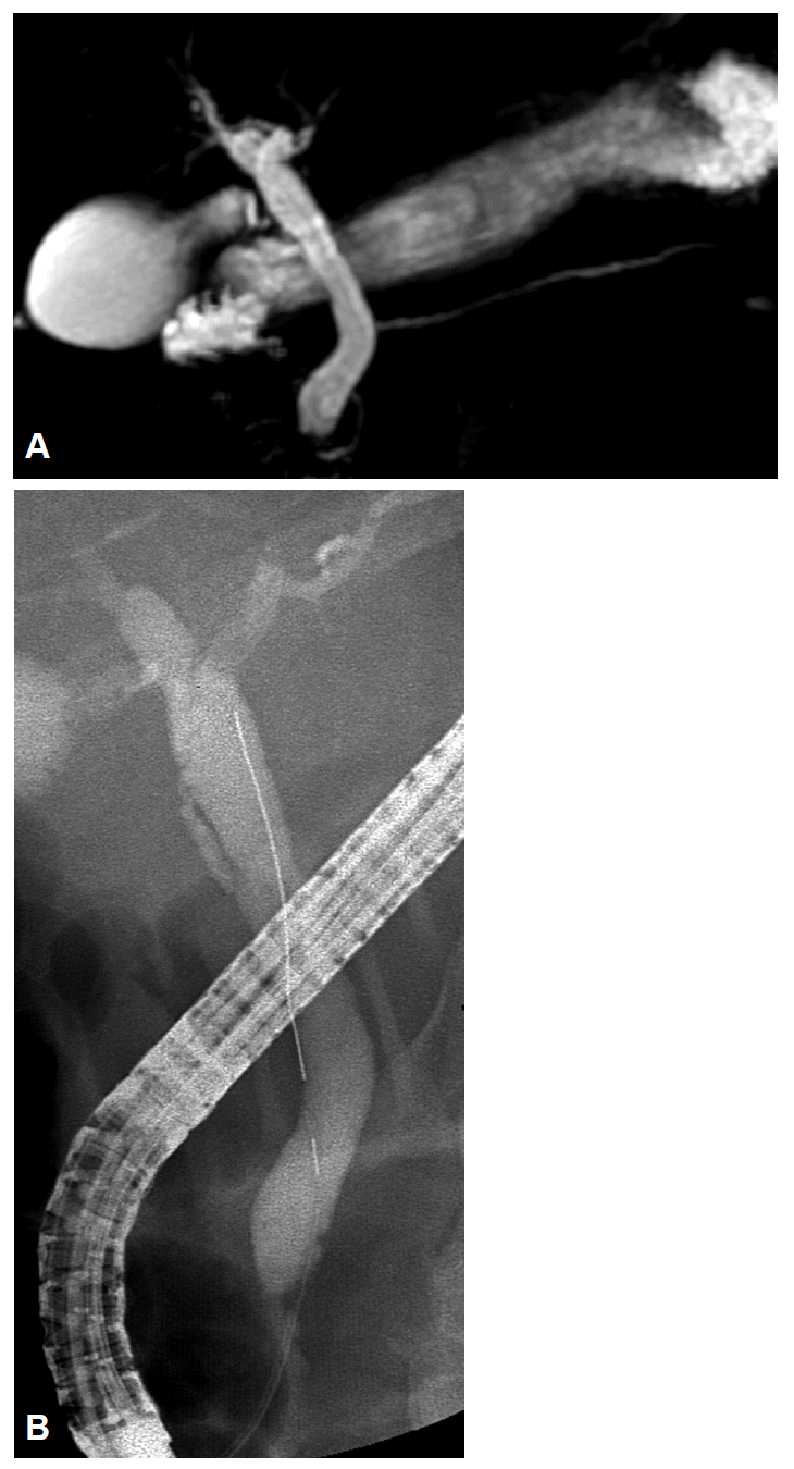
(A) Magnetic resonance cholangiopancreatography showing dilated extrahepatic bile duct without any definite cause of obstruction. (B) Endoscopic retrograde cholangiogram does not reveal any cause of acute cholangitis.

(A) Initial findings of the intraductal ultrasonography revealing crescent-layered wall thickening in the right main hepatic duct. (B) Intraductal ultrasonography showing an actively motile tubular structure in the bile duct. (C) Endoscopic appearance of Fasciola hepatica showing a leaf-like trematode extracted by using a balloon catheter. (D) Finding of F. hepatica after fixation in formalin.
DISCUSSION
F. hepatica is a zoonotic trematode that infects sheep, goats, and cattle as normal hosts. Humans acquire the parasite after ingestion of aquatic plants, such as watercress, that are infested with metacercariae, drinking contaminated water, or eating the uncooked liver of infested animals [1]. Human fascioliasis has two stages: the acute hepatic stage and the chronic biliary stage. The infected metacercariae excyst in the small bowel, penetrate the intestinal wall, migrate to the liver, and penetrate the liver capsule and hepatic parenchyma to the bile duct. The flukes then reside within the bile ducts. In its chronic biliary phase, F. hepatica infestation results in periodic bile duct obstruction, inducing obstructive jaundice, cholangitis, and/or acute biliary pancreatitis [1,6,9,10]. A diagnosis of human fascioliasis is usually achieved by detecting eggs in the stool or a serological test for antibodies against the worm. However, these diagnostic approaches may not be practical without suspicion for fascioliasis; especially in non-endemic areas [1,2].
In non-endemic areas, diagnostic evaluation in patients with acute cholangitis and biliary pancreatitis is typically performed from the perspective of choledocholithiasis or biliary malignancy, which are more common diseases. Indeed, vague findings of bile duct obstruction by F. hepatica may lead to a clinical misdiagnosis of cholangiocarcinoma in imaging studies, including computed tomography and/or MRCP [11-13]. For human fascioliasis, ERCP should be performed to evaluate and treat bile duct obstruction that allows the detection and extraction of the motile leaf-like worm [3,4,7]. In previous reports, ERCP had a key role in the diagnosis and treatment of biliary fascioliasis. However, a cholangiogram did not reveal the cause of the cholangitis in this case. The leaf-like structure of F. hepatica enables its attachment to the bile duct wall, and can be overlooked on a cholangiogram. IDUS is readily performed without a sphincterotomy during ERCP and is a very sensitive imaging modality for the bile duct. It is also useful for the differential diagnosis of obstructive jaundice in patients with negative cholangiogram findings. Kim et al. [14] reported the use of IDUS in patients with highly suspected choledocholithiasis but normal ERCP findings. IDUS can detect occult small stones and sludges that would otherwise not be revealed in imaging studies, including ERCP. We performed IDUS to investigate the cause of bile duct obstruction and detected a motile tubular structure. Even in the IDUS evaluation, the leaf-like worm attached to the bile duct was confused as thickening of the bile duct wall. However, another advantage of IDUS is the provision real-time images, and the live motile worm was detected using IDUS. EUS can also be a sensitive imaging modality for the extrahepatic bile duct in real-time, and may be useful for the diagnosis of biliary fascioliasis [8]. However, EUS may be limited for evaluation of the proximal or intrahepatic bile duct. In this case, IDUS revealed the worm in the proximal bile duct. The detected biliary fascioliasis could therefore be treated during the same endoscopic session without endoscope exchange because IDUS is performed adjunctively during ERCP.
In conclusion, F. hepatica induced acute cholangitis in this case. However, it was not detected in imaging studies, including a cholangiogram. Wire-guided IDUS revealed an actively moving worm in the bile duct. IDUS findings of F. hepatica in the bile duct have not been reported previously. Thus, we present this apparently unique case of human fascioliasis diagnosed using IDUS.
Notes
Conflicts of Interest: The authors have no financial conflicts of interest.
Acknowledgements
This article was supported partially by the Soonchunhyang University Research Fund.