INTRODUCTION
Gastrointestinal tract duplications are uncommon congenital anomalies and found anywhere along the alimentary tract from the mouth to the anus. Especially, esophageal duplication (ED) is rarely diagnosed in adults. The estimated incidence of congenital ED is 1:8,200, with a male sex predominance [1,2]. Almost all of the reported cases were detected with respiratory distress manifestation in the early days of life. Most EDs are found in the distal third of the esophagus and are frequently incidental findings on routine chest radiography. ED is divided into three types: cystic (the most common type), tubular, and diverticular [3]. ED can be associated with other congenital anomalies such as small intestine duplications, esophageal atresia, bronchoesophageal fistulas (BEFs), and spinal abnormalities [4]. Less than 20% of EDs have communication with the main lumen through a tubular tract [5]. Also, ED could communicate with the tracheobronchial tree and create fistulae.
Communicating tubular ED is extremely rare, and only one case report of communicating tubular ED combined with BEF in an adult has been published in the Japanese literature (in 1994) [6]. There has been no report of such a case in the English literature.
Here, we report a case of communicating tubular ED combined with BEF, diagnosed on the basis of endoscopy and esophagography findings during the postoperational evaluation of BEF.
CASE REPORT
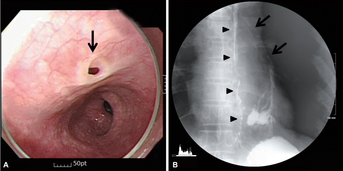
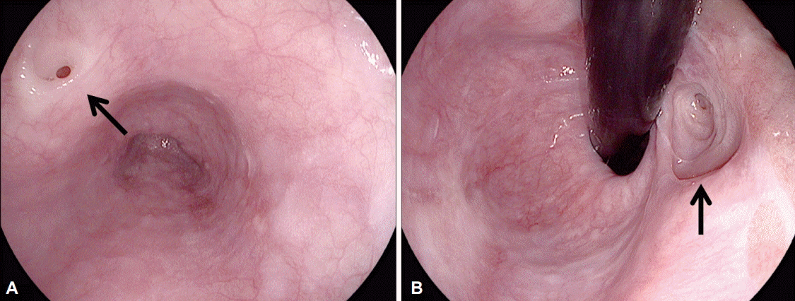
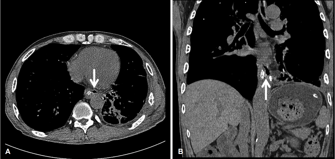
A 49-year-old male patient was admitted to our hospitalтs emergency center because of herbicide poisoning. After drinking several glasses of alcohol and herbicide, he presented with persistent nausea and vomiting. He had been drinking about 54 g alcohol daily and had 50-pack-year history of cigarette smoking. He had a history of pulmonary tuberculosis and received antituberculosis medication 8 years ago. At that time, he was also found to have BEF and alcoholic liver disease. Intermittent and persistent cough had developed for a while. On admission, initial physical examination revealed no specific finding except for an elevated respiratory rate of 28/min. The results of laboratory tests were as follows: white blood cell count 15,660/mm3 , hemoglobin 12.9 g/dL, hematocrit 38.1%, platelet count 155,000/mm3 , total protein 7.9 g/dL, albumin 3.7 g/dL, aspartate aminotransferase 80 U/L, alanine aminotransferase 37 U/L, prothrombin time 1.03, and C-reactive protein 3.48 mg/dL. The results of blood gas analysis were pH 7.513, pCO2 35.2 mm Hg, pO2 80.5 mm Hg, and HCO3 27.7 mmol/L. Chest radiography demonstrated a consolidation in the lower left zone of the lungs. After aspiration pneumonia was diagnosed, the patient was treated with conservative management. On the second hospital day, he developed severe persistent cough. On endoscopic and bronchoscopic examinations, a fistula opening was found at the mid-esophagus, and the opening had a whitish surface with a slightly screwed pattern without inflammatory sign or discharge (Fig. 1A). We could not localize the other opening or a fistula tract. A previous esophagography image (Fig. 1B) taken at a local hospital revealed a communicating fistula tract between the bronchus and the lower esophagus (BEF); however, a chest computed tomography (CT) scan, also taken at the local hospital, did not show discrete BEF. On the 6th hospital day, his pneumonic condition was well improved and he could ambulate. On the 10th hospital day, he underwent surgical operation for the repair of the BEF. During the operation, a 2-cm-long fistula tract between the lower esophagus and the medial basal segment of the left lung was noted, which was successfully removed and repaired. His condition was stable, and he had no complication. On the 3rd postoperational day, a follow-up esophagography for the postoperational evaluation revealed contrast leakage at the left side of the mid-esophagus and drainage to the distal esophagus (Fig. 2). We thought that the leakage resulted from an unidentified condition, such as ED with esophago-esophageal fistula (Fig. 2). On a follow-up endoscopic examination, a proximal fistula opening was found at the mid-esophagus and the distal opening was found on the cardia of the stomach, located in the hiatal hernia (Fig. 3). On chest CT, an about 7-mm air-filled tract was noted, one end of which was connected to the distal esophagus and the other end was connected to the cardia of the stomach (Fig. 4). The final diagnosis was communicating tubular ED with BEF (Fig. 5). Initially, he was operated for the symptomatic BEF; however, a communicating ED was later detected incidentally during a postoperational follow-up. The symptoms of BEF were improved without any unusual complication, and he wanted to be discharged without any further treatment or surgery. At 2 years of follow-up, he has been doing well without any complication of ED with BEF.
DISCUSSION
The only case of a communicating tubular ED with BEF was published in the Japanese literature. The patient was a 51-year-old woman who chronically experienced coughing when drinking water. Esophagoscopy revealed tubular ED with BEF, and endoscopy showed one small opening of ED. She was operated to remove the BEF with ED [6].
Most EDs are found in the distal third of the esophagus and are frequently asymptomatic; however, dysphagia, respiratory distress, recurrent pneumonia, vomiting, failure to thrive, gastroesophageal reflux, melena, and anemia may also be present in rare cases [1,7].
At the time of admission, our patient had chronic intermittent cough and knew about his BEF. Pneumonia might be considered a complication of drug intoxication or BEF. On admission, persistent cough developed abruptly, and he wanted to undergo evaluation and receive treatment for his disease. Chest CT and endoscopic examination did not reveal a communicating ED at that time; however, postoperational follow-up esophagography for the confirmation of fistula closure revealed another remaining fistula tract. This fistula tract communicated not with the bronchus but with the distal esophagus as a tubular structure, which is an evidence of the possibility of ED. Therefore, endoscopic reexamination and follow-up chest CT were done, and we finally confirmed ED.
Later, we incidentally found that he had a communicating tubular ED with BEF, which we could not find on the initial evaluation. The ED was initially not detected because we thought the cough symptom was caused by the BEF that was detected on esophagography at the previous hospital. Another possible reason is that the ED was not obvious on the esophagography. The ED was congenitally accompanied by the BEF, and they shared the same opening, which might have caused the relatively less influx of the contrast medium into the ED than into the BEF, making the ED undetectable on esophagography. According to the assumption from interdisciplinary discussion, it is highly likely that the removal of the BEF might have made the ED easily accessible for the contrast medium and thus more detectable on esophagography. This is also supported by the fact that ED was suspected on the review of the previous CT images (images not presented here).
One major limitation of this case report is that the possibility of an esophagogastric fistula, not an ED, could not be confirmed by histology. In another report with a similar case to ours, ED was verified on the basis of the histological findings [8]. However, as the endoscopic findings suggest (Fig. 3B), the orifice inside the hiatal hernia appeared to be covered by the epithelium of the esophagus, suggesting the possibility of an orifice from the ED, rather than an esophagogastric fistula. A histological examination would be helpful for differential diagnosis in patients showing similar findings in the future.
Identifying an ED is very difficult. Upper gastrointestinal endoscopy can help in the diagnosis through direct vision; however, it cannot determine whether the ED is a communicating or a noncommunicating type. In our case, the initial endoscopic examination for the confirmation of BEF could not find a communicating tubular ED. However, the result of the follow-up esophagography revealed the possibility of a communicating tubular ED, and we conducted repeat endoscopy carefully. The follow-up endoscopy found two openings of ED. As can be seen in Fig. 3, the distal orifice of the ED was at the sliding hiatal hernia sac, and it was very difficult to find. The opening could be seen along the whole esophagus as a whitish or pinkish hole not discriminated from the surrounding opening tissue.
Several imaging studies for diagnostic workup were also needed. Radiological methods can be helpful in the diagnosis and localization of the disease. Esophagography may show displacement of the esophagus due to a paraesophageal mass; however, a tubular ED may not influence the esophagus [9]. Contrast esophagographic evaluation could be useful in diagnosing a communicating ED preoperatively, and to confirm that a leak is not present during the postoperative follow-up period. CT could demonstrate the exact anatomic position of the fistular tract and influence the decision making about resectability. Magnetic resonance imaging, endoscopic ultrasonography, and Tc-99m pertechnetate scintigraphy could be helpful for the diagnosis of this disease [8]. However, these methods yield mainly inconclusive results [10,11].
The management of ED is dependent on the type and size of the ED, and the severity of the symptoms. Complete resection is a well-documented treatment for duplication of the esophagus; however, conservative management can be an option for nonsymptomatic patients. Symptomatic ED has mainly been managed with extensive surgery through a thoracotomy [11]. With recent advances in minimally invasive surgery, surgical treatment has improved in efficacy and offers advantages to patients. A clear exposure and identification of the duplication are important for a successful operation. After surgical excision, the prognosis is encouraging. As our patient received primary repair of BEF without knowledge of ED, and his symptoms disappeared, we did not conduct additional surgery for ED. After surgery, he has been doing well during the follow-up period.
In conclusion, identifying a communicating ED with BEF is difficult. On the diagnostic examination for a BEF, the possibility of ED should be considered and careful endoscopic examination is also important for the detection of fistula orifices.


