Skip Navigation
Skip to contents

Clin Endosc : Clinical Endoscopy

OPEN ACCESS

Articles

Page Path
HOME > Clin Endosc > Volume 49(2); 2016 > Article
Brief Report Laterally Spreading Tumor of the Rectum Delineated with Linked Color Imaging Technology
Masahiro Okada1, Hirotsugu Sakamoto1, Takahito Takezawa1, Yoshikazu Hayashi1, Keijiro Sunada1, Alan K. Lefor2, Hironori Yamamoto1,
Clinical Endoscopy 2016;49(2):207-208.
DOI: https://doi.org/10.5946/ce.2015.077
Published online: February 12, 2016

1Division of Gastroenterology, Department of Medicine, Jichi Medical University, Shimotsuke, Japan

2Department of Surgery, Jichi Medical University, Shimotsuke, Japan

Correspondence: Hironori Yamamoto Division of Gastroenterology, Department of Medicine, Jichi Medical University, 3311-1 Yakushiji, Shimotsuke 329-0498, Japan Tel: +81-285-58-7347, Fax: +81-285-40-6598, E-mail: ireef@jichi.ac.jp
• Received: May 21, 2015   • Revised: June 12, 2015   • Accepted: June 14, 2015

Copyright © 2016 Korean Society of Gastrointestinal Endoscopy

This is an Open Access article distributed under the terms of the Creative Commons Attribution Non-Commercial License (http://creativecommons.org/licenses/by-nc/3.0/) which permits unrestricted non-commercial use, distribution, and reproduction in any medium, provided the original work is properly cited.

  • 9,367 Views
  • 156 Download
  • 38 Web of Science
  • 37 Crossref
  • 41 Scopus
prev
Colonoscopy has been shown to decrease the incidence of colorectal cancer and cancer death through the removal of adenomatous polyps. To make an accurate diagnosis of colorectal neoplasms, several image-enhanced endoscopic systems, such as narrow band imaging (NBI), flexible spectral imaging color enhancement, and blue laser imaging (BLI), have been developed. However, no significant differences have been reported in the adenoma detection rate between image-enhanced endoscopy and conventional white light endoscopy [1]. NBI and BLI are often criticized for darkening the endoscopy image and, in turn, hampering a wider view of the colon [2]. Moreover, the miss rate for flat lesions was significantly higher than that for pedunculated lesions [3]. We present a case that suggests a possible improvement in adenoma detection rates, especially for flat adenomas.
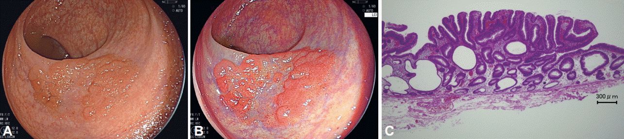
A 66-year-old woman with a 40-mm laterally spreading tumor of the rectum was referred for endoscopic resection. Before the endoscopic resection, we observed the lesion with an EC-L590ZW endoscope with the LASEREO system (FUJIFILM Co., Tokyo, Japan), with white light (Fig. 1A) and with Linked Color Imaging (LCI) (Fig. 1B). The lesion was clearly seen as a bright reddish area on LCI. Compared with the white light image, the LCI image makes the lesion more easily recognizable, thanks to the striking color contrast between the neoplastic mucosa and the normal mucosa. The lesion was diagnosed as an adenoma on the basis of the magnified BLI images. Successful en bloc endoscopic submucosal dissection was performed for the lesion, and the diagnosis of high-grade adenoma with negative resection margins was confirmed histopathologically (Fig. 1C).
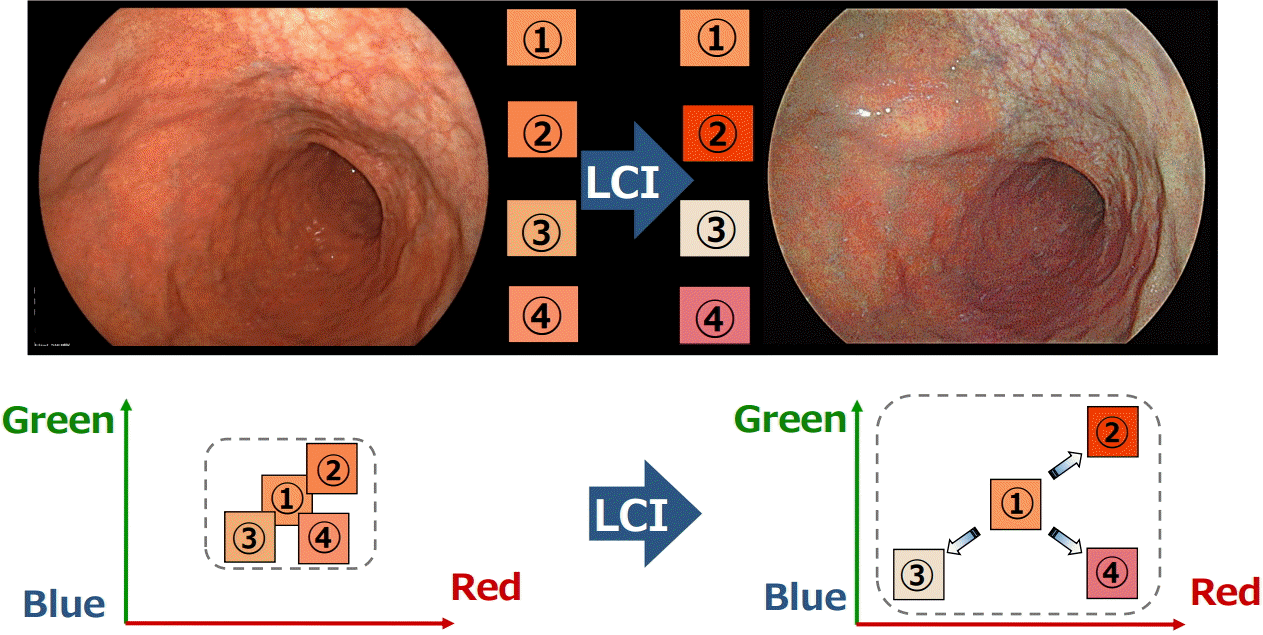
NBI is the most widely used system among several available image enhanced endoscopy systems. NBI modifies the center wavelength and bandwidth of an endoscope’s light into narrow-band illumination at 415±30 nm within the hemoglobin absorption band, thereby facilitating a clearer visualization of vascular structures [4]. However, this technique has limitations such as dark imaging of distant lesions because of narrow-band illumination. BLI was developed to compensate for these inherent limitations of NBI. BLI uses narrow-band laser light combined with white light. This combination results in a bright image of the digestive mucosa, enabling the detailed visualization of both the microstructure and the microvasculature [5]. However, BLI still is not able to obtain sufficient brightness for distant lesions. The newly developed LCI system (FUJIFILM Co.) creates clear and bright endoscopic images by using short-wavelength narrow-band laser light combined with white laser light on the basis of BLI technology. This system can obtain bright endoscopic images even at a distant view because LCI has more intense white light than the short-wavelength narrow-band laser light. Short-wavelength narrow-band laser light enhances the vessels on the mucosal surface and the patterns of the mucosa, which means that BLI enables a clearer visualization of microvascular structures than does LCI. In contrast, LCI enhances differences in hue, in the red region of the spectrum, through digital processing (Fig. 2). This makes red areas appear redder and white areas appear whiter. Thus, it is easier to recognize a slight difference in color of the mucosa. Therefore, LCI may facilitate the detection of flat colorectal neoplasms without magnification. After the detection of lesions, BLI images are easily produced with a push of a button for use in qualitative diagnosis. Further studies are needed to confirm the utility of LCI.
Fig. 1.
Endoscopic images of a laterally spreading tumor (LST). (A) White light endoscopic view of an LST, 40 mm in diameter, in the rectum. (B) The lesion is easily recognizable as a bright reddish area on Linked Color Imaging. (C) Pathological image of the resected lesion (H&E stain, ×4).
ce-2015-077f1.gif
Fig. 2.
Principles of Linked Color Imaging (LCI). LCI enhances differences in hue, in the red region of the spectrum, through digital processing.
ce-2015-077f2.gif
  • 1. Kamiński MF, Hassan C, Bisschops R, et al. Advanced imaging for detection and differentiation of colorectal neoplasia: European Society of Gastrointestinal Endoscopy (ESGE) Guideline. Endoscopy 2014;46:435–449.ArticlePubMedPDF
  • 2. Dinesen L, Chua TJ, Kaffes AJ. Meta-analysis of narrow-band imaging versus conventional colonoscopy for adenoma detection. Gastrointest Endosc 2012;75:604–611.ArticlePubMed
  • 3. Heresbach D, Barrioz T, Lapalus MG, et al. Miss rate for colorectal neoplastic polyps: a prospective multicenter study of back-to-back video colonoscopies. Endoscopy 2008;40:284–290.ArticlePubMed
  • 4. Emura F, Saito Y, Ikematsu H. Narrow-band imaging optical chromocolonoscopy: advantages and limitations. World J Gastroenterol 2008;14:4867–4872.ArticlePubMedPMC
  • 5. Osawa H, Yamamoto H. Present and future status of flexible spectral imaging color enhancement and blue laser imaging technology. Dig Endosc 2014;26 Suppl 1:105–115.ArticlePubMed

Figure & Data

REFERENCES

    Citations

    Citations to this article as recorded by  
    • Significance of Endoscopic Redness of Duodenum in Health Checkup
      Atsushi Nakayama, Motohiko Kato, Yoshiki Sakaguchi, Yu Takahashi, Shinya Kodashima, Ai Fujimoto, Nobutake Yamamichi, Kazumasa Miki, Naohisa Yahagi
      JGH Open.2025;[Epub]     CrossRef
    • Practical utility of linked color imaging in colonoscopy: Updated literature review
      Fumiaki Ishibashi, Sho Suzuki
      Digestive Endoscopy.2024;[Epub]     CrossRef
    • The Past, Present and Future of Imaging Enhanced Endoscopy in Colon Tumor
      Kyueng-Whan Min, One-Zoong Kim
      Journal of Digestive Cancer Research.2024; 12(2): 90.     CrossRef
    • Linked color imaging and upper gastrointestinal neoplasia
      Shoko Ono, Masaki Inoue, Masayuki Higashino, Shuhei Hayasaka, Shugo Tanaka, Hiroki Egami, Naoya Sakamoto
      Digestive Endoscopy.2024;[Epub]     CrossRef
    • Linked color imaging improves the diagnostic accuracy of eosinophilic esophagitis
      Yasuhiko Abe, Yu Sasaki, Makoto Yagi, Naoko Mizumoto, Yusuke Onozato, Takashi Kon, Masakuni Shoji, Kazuhiro Sakuta, Takayuki Sakai, Matsuki Umehara, Minami Ito, Shuhei Nakamura, Hidemoto Tsuchida, Yoshiyuki Ueno
      DEN Open.2023;[Epub]     CrossRef
    • Improvement of detection sensitivity of upper gastrointestinal epithelial neoplasia in linked color imaging based on data of eye tracking
      Masayuki Higashino, Shoko Ono, Shogo Matsumoto, Marina Kubo, Naohiro Yasuura, Shuhei Hayasaka, Ikko Tanaka, Yoshihiko Shimoda, Yusuke Nishimura, Masayoshi Ono, Keiko Yamamoto, Yuji Ono, Naoya Sakamoto
      Journal of Gastroenterology and Hepatology.2023; 38(5): 710.     CrossRef
    • Efficacy of image-enhanced endoscopy for colorectal adenoma detection: A multicenter, randomized trial
      Zhi-Peng Qi, En-Pan Xu, Dong-Li He, Yan Wang, Bai-Sheng Chen, Xue-Si Dong, Qiang Shi, Shi-Lun Cai, Qi Guo, Ni Li, Xing Li, Hai-Yan Huang, Bing Li, Di Sun, Jian-Guang Xu, Zhang-Han Chen, Ayimukedisi Yalikong, Jin-Yi Liu, Zhen-Tao Lv, Jian-Min Xu, Ping-Hong
      World Journal of Gastrointestinal Oncology.2023; 15(5): 878.     CrossRef
    • Using the string‐clip method to retrieve the resected specimen allowed a clear observation of the colon and detection of a new lesion
      Xinghua Ma, Kazumasa Kawashima, Yutaka Saito
      Digestive Endoscopy.2022;[Epub]     CrossRef
    • Linked color imaging improves identification of early gastric cancer lesions by expert and non-expert endoscopists
      Kiki Fockens, Jeroen de Groof, Joost van der Putten, Tsevelnorov Khurelbaatar, Hisashi Fukuda, Takahito Takezawa, Yoshimasa Miura, Hiroyuki Osawa, Hironori Yamamoto, Jacques Bergman
      Surgical Endoscopy.2022; 36(11): 8316.     CrossRef
    • Diagnostic accuracy of linked colour imaging versus white light imaging for early gastric cancers: a prospective, multicentre, randomized controlled trial study
      Min Min, Xiaotian Sun, Jianying Bai, Qinsheng Zhang, Xiaocui Yang, Qiang Guo, Rong Wang, Bangmao Wang, Zhiwu Lv, Jie Pan, Chunmeng Jiang, Duanmin Hu, Bing Nong, Enqiang Linghu, Yan Liu
      Annals of Medicine.2022; 54(1): 3305.     CrossRef
    • A case of Cronkhite–Canada syndrome with repeated linked color imaging observation of the subepithelial capillary network in the colon
      Yayoi Matsumoto, Fumikazu Koyama, Kohei Morita, Hiroyuki Kuge, Shinsaku Obara, Yosuke Iwasa, Takeshi Takei, Tomomi Sadamitsu, Chiho Ohbayashi, Masayuki Sho
      Clinical Journal of Gastroenterology.2022; 15(5): 934.     CrossRef
    • Linked color imaging improves visibility of colorectal serrated lesion by high color contrast to surrounding mucosa
      Takashi Murakami, Eiji Kamba, Kei Nomura, Taro Kurosawa, Keiichi Haga, Hirofumi Fukushima, Tsutomu Takeda, Tomoyoshi Shibuya, Takashi Yao, Akihito Nagahara
      Digestive Endoscopy.2022; 34(7): 1422.     CrossRef
    • Using texture and colour enhancement imaging to evaluate gastrointestinal diseases in clinical practice: a review
      Mitsushige Sugimoto, Yohei Koyama, Takao Itoi, Takashi Kawai
      Annals of Medicine.2022; 54(1): 3314.     CrossRef
    • Gastric intestinal metaplasia assessment between linked color imaging based on endoscopy and pathology
      Guanpo Zhang, Jin Zheng, Linfu Zheng, Shentong Yu, Chuanshen Jiang, Wulian Lin, Dazhou Li, Lijuan Qu, Wen Wang
      Scandinavian Journal of Gastroenterology.2021; 56(1): 103.     CrossRef
    • Optical effect of spraying l-menthol on gastric intestinal metaplasia visualized by linked color imaging
      Shoko Ono, Yoshihiko Shimoda, Ikko Tanaka, Sayoko Kinowaki, Masaki Inoue, Masayoshi Ono, Keiko Yamamoto, Yuichi Shimizu, Naoya Sakamoto
      European Journal of Gastroenterology & Hepatology.2021; 33(3): 358.     CrossRef
    • Linked colour imaging versus white‐light colonoscopy for the detection of flat colorectal lesions: A randomized controlled trial
      Takahiro Kudo, Akira Horiuchi, Reiko Kyodo, Ichitaro Horiuchi, Nobuyasu Arai, Masashi Kajiyama, Naoki Tanaka
      Colorectal Disease.2021; 23(6): 1414.     CrossRef
    • Application of linked color imaging in the diagnosis of early gastrointestinal neoplasms and precancerous lesions: a review
      Shanshan Wang, Lei Shen, Hesheng Luo
      Therapeutic Advances in Gastroenterology.2021;[Epub]     CrossRef
    • Linked-color Imaging May Help Improve the Visibility of Superficial Barrett's Esophageal Adenocarcinoma by Increasing the Color Difference
      Masahiro Saito, Tomoyuki Koike, Yuki Ohara, Kenichiro Nakagawa, Takeshi Kanno, Xiaoyi Jin, Waku Hatta, Kaname Uno, Naoki Asano, Akira Imatani, Atsushi Masamune
      Internal Medicine.2021; 60(21): 3351.     CrossRef
    • Colon polyp detection using linked color imaging compared to white light imaging: Systematic review and meta‐analysis
      Satoshi Shinozaki, Yasutoshi Kobayashi, Yoshikazu Hayashi, Hirotsugu Sakamoto, Keijiro Sunada, Alan Kawarai Lefor, Hironori Yamamoto
      Digestive Endoscopy.2020; 32(6): 874.     CrossRef
    • Significance of Linked Color Imaging for Predicting the Risk of Clinical Relapse in Ulcerative Colitis
      Shuji Kanmura, Akihito Tanaka, Kazuki Yutsudou, Kosuke Kuwazuru, Fukiko Komaki, Yuga Komaki, Hiromichi Iwaya, Shiho Arima, Fumisato Sasaki, Shiroh Tanoue, Shinichi Hashimoto, Akio Ido
      Gastroenterology Research and Practice.2020; 2020: 1.     CrossRef
    • Linked color imaging can enhance recognition of early gastric cancer by high color contrast to surrounding gastric intestinal metaplasia
      Hisashi Fukuda, Yoshimasa Miura, Hiroyuki Osawa, Takahito Takezawa, Yuji Ino, Masahiro Okada, Tsevelnorov Khurelbaatar, Alan Kawarai Lefor, Hironori Yamamoto
      Journal of Gastroenterology.2019; 54(5): 396.     CrossRef
    • Advanced Treatment and Imaging in Colonoscopy: The Pocket-Creation Method for Complete Resection and Linked Color Imaging for Better Detection of Early Neoplastic Lesions by Colonoscopy
      Hironori Yamamoto, Satoshi Shinozaki, Yoshikazu Hayashi, Yoshimasa Miura, Tsevelnorov Khurelbaatar, Hiroyuki Osawa, Alan Kawarai Lefor
      Clinical Endoscopy.2019; 52(2): 107.     CrossRef
    • Objective evaluation of the visibility of colorectal lesions using eye tracking
      Kana Kumahara, Hiroaki Ikematsu, Kensuke Shinmura, Tatsuro Murano, Atsushi Inaba, Kei Okumura, Keiichiro Nishihara, Hironori Sunakawa, Yasuaki Furue, Renma Ito, Daiki Sato, Tatsunori Minamide, Naoki Okamoto, Yoichi Yamamoto, Masayuki Suyama, Kenji Takashi
      Digestive Endoscopy.2019; 31(5): 552.     CrossRef
    • Blue Laser Imaging with a Small-Caliber Endoscope Facilitates Detection of Early Gastric Cancer
      Haruo Takahashi, Yoshimasa Miura, Hiroyuki Osawa, Takahito Takezawa, Yuji Ino, Masahiro Okada, Alan Kawarai Lefor, Hironori Yamamoto
      Clinical Endoscopy.2019; 52(3): 273.     CrossRef
    • Linked color imaging for the detection of early gastrointestinal neoplasms
      Satoshi Shinozaki, Hiroyuki Osawa, Yoshikazu Hayashi, Alan Kawarai Lefor, Hironori Yamamoto
      Therapeutic Advances in Gastroenterology.2019;[Epub]     CrossRef
    • Evaluation of the impact of linked color imaging for improving the visibility of colonic polyp
      Yasuyoshi Tanaka, Takuya Inoue, Kazuki Kakimoto, Kei Nakazawa, Hideki Tawa, Yuki Hirata, Toshihiko Okada, Sadaharu Nouda, Ken Kawakami, Toshihisa Takeuchi, Yutaro Egashira, Kazuhide Higuchi
      Oncology Letters.2019;[Epub]     CrossRef
    • Linked color imaging improves detection of minimal change esophagitis in non-erosive reflux esophagitis patients
      Pei Deng, Min Min, Tenghui Dong, Yiliang Bi, Airong Tang, Yan Liu
      Endoscopy International Open.2018; 06(10): E1177.     CrossRef
    • Linked color imaging enhances gastric cancer in gastric intestinal metaplasia
      Shoko Ono, Satoshi Abiko, Mototsugu Kato
      Digestive Endoscopy.2017; 29(2): 230.     CrossRef
    • Flexible Gastro-intestinal Endoscopy — Clinical Challenges and Technical Achievements
      Niehls Kurniawan, Martin Keuchel
      Computational and Structural Biotechnology Journal.2017; 15: 168.     CrossRef
    • Comparison of linked color imaging and white-light colonoscopy for detection of colorectal polyps: a multicenter, randomized, crossover trial
      Min Min, Pei Deng, Wenhua Zhang, Xiaomin Sun, Yan Liu, Bing Nong
      Gastrointestinal Endoscopy.2017; 86(4): 724.     CrossRef
    • Linked colour imaging benefits the endoscopic diagnosis of distal gastric diseases
      Xiaotian Sun, Yiliang Bi, Tenghui Dong, Min Min, Wei Shen, Yang Xu, Yan Liu
      Scientific Reports.2017;[Epub]     CrossRef
    • Image-enhanced endoscopy: How far do we need to go?
      Hazem Hammad, Tonya Kaltenbach, Roy Soetikno
      Gastrointestinal Endoscopy.2017; 86(4): 698.     CrossRef
    • Objective Endoscopic Analysis with Linked Color Imaging regarding Gastric Mucosal Atrophy: A Pilot Study
      Kazuhiro Mizukami, Ryo Ogawa, Kazuhisa Okamoto, Mitsutaka Shuto, Kensuke Fukuda, Akira Sonoda, Osamu Matsunari, Yuka Hirashita, Tadayoshi Okimoto, Masaaki Kodama, Kazunari Murakami
      Gastroenterology Research and Practice.2017; 2017: 1.     CrossRef
    • 3) Progress in Endoscopy
      Hironori Yamamoto
      Nihon Naika Gakkai Zasshi.2017; 106(9): 1796.     CrossRef
    • Endoscopic Submucosal Dissection of a Rectal Neuroendocrine Tumor Using Linked Color Imaging Technique
      Xiao-Tian Sun, Min Min, Yi-Liang Bi, Yang Xu, Yan Liu
      Chinese Medical Journal.2017; 130(9): 1127.     CrossRef
    • Linked color imaging application for improving the endoscopic diagnosis accuracy: a pilot study
      Xiaotian Sun, Tenghui Dong, Yiliang Bi, Min Min, Wei Shen, Yang Xu, Yan Liu
      Scientific Reports.2016;[Epub]     CrossRef
    • Linked color imaging technology facilitates early detection of flat gastric cancers
      Hisashi Fukuda, Yoshimasa Miura, Yoshikazu Hayashi, Takahito Takezawa, Yuji Ino, Masahiro Okada, Hiroyuki Osawa, Alan K. Lefor, Hironori Yamamoto
      Clinical Journal of Gastroenterology.2015; 8(6): 385.     CrossRef

    • PubReader PubReader
    • ePub LinkePub Link
    • Cite
      CITE
      export Copy Download
      Close
      Download Citation
      Download a citation file in RIS format that can be imported by all major citation management software, including EndNote, ProCite, RefWorks, and Reference Manager.

      Format:
      • RIS — For EndNote, ProCite, RefWorks, and most other reference management software
      • BibTeX — For JabRef, BibDesk, and other BibTeX-specific software
      Include:
      • Citation for the content below
      Laterally Spreading Tumor of the Rectum Delineated with Linked Color Imaging Technology
      Clin Endosc. 2016;49(2):207-208.   Published online February 12, 2016
      Close
    • XML DownloadXML Download
    Figure
    • 0
    • 1
    Laterally Spreading Tumor of the Rectum Delineated with Linked Color Imaging Technology
    Image Image
    Fig. 1. Endoscopic images of a laterally spreading tumor (LST). (A) White light endoscopic view of an LST, 40 mm in diameter, in the rectum. (B) The lesion is easily recognizable as a bright reddish area on Linked Color Imaging. (C) Pathological image of the resected lesion (H&E stain, ×4).
    Fig. 2. Principles of Linked Color Imaging (LCI). LCI enhances differences in hue, in the red region of the spectrum, through digital processing.
    Laterally Spreading Tumor of the Rectum Delineated with Linked Color Imaging Technology

    Clin Endosc : Clinical Endoscopy Twitter Facebook
    Close layer
    TOP