An Intractable Caustic Esophageal Stricture Successfully Managed with Sequential Treatment Comprising Incision with an Insulated-Tip Knife, Balloon Dilation, and an Oral Steroid
Article information
Abstract
Bougie or balloon dilation is a good short-term treatment for caustic esophageal strictures, although recurrence after dilation occurs in approximately 30% of these cases. Therefore, long-term treatment options are required in some cases, and endoscopic incisional therapy has been used for patients with an anastomotic stricture in the gastrointestinal tract. A 58-year-old woman presented with severe swallowing difficulty because of a caustic esophageal stricture, which was caused by accidental exposure to anhydrous acetic acid at infancy. She had undergone several previous bougie and balloon dilations but the stricture did not improve. We performed sequential treatment comprising incision with an insulated-tip knife, balloon dilation, and an oral steroid, which resulted in the patient’s symptoms markedly improving. Thus, we report this case of an intractable caustic esophageal stricture, which was successfully treated using combined endoscopic sequential treatment.
INTRODUCTION
Exposure of the esophagus to caustic material can lead to necrosis, ulceration, fibrosis, and ultimately stricture. Balloon and bougie dilation have similar efficacies for treating these strictures [1,2], although stricture recurrence after balloon dilation can occur in up to 30% of these cases [3,4]. Furthermore, a long-term response cannot be guaranteed in cases of non-peptic strictures [5], and symptomatic recurrence frequently occurs within 1 year after the initial dilation. Thus, caustic esophageal strictures occasionally require a long-term treatment option, and one case report described endoscopic incisional therapy for a patient with a refractory caustic esophageal stricture [6]. We report a case of an intractable caustic esophageal stricture that was successfully managed with sequential treatment that comprised incision with an insulated-tip knife, balloon dilation, and an oral steroid.
CASE REPORT
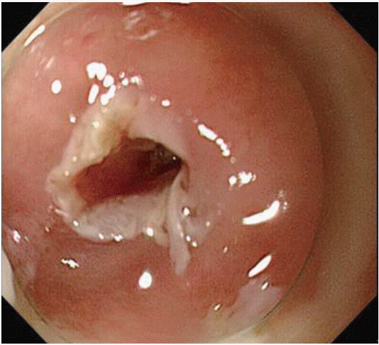
A 58-year-old woman presented with severe swallowing difficulty, because of an esophageal stricture that was caused by accidental exposure to anhydrous acetic acid at infancy. The patient reported that she had to manually compress the site of the stricture to swallow solid foods, such as meat, and she had grade 2 dysphagia [7]. The esophageal stricture was located 18 cm from the incisor (Fig. 1). She was originally treated using bougie dilation 22 years before the present admission, and we had attempted four balloon dilations during the last 10 years, although the esophageal stricture remained intractable (Figs. 2, 3). Therefore, we performed incisional therapy parallel to the longitudinal axis of the esophagus using an insulated-tip electrosurgical knife (ITknife nano, KD-612L/U; Olympus, Tokyo, Japan). The electrical current was generated using an ICC200 unit (ERBE Elektromedizin GmbH, Tubingen, Germany), which was set to the pure cut current (endocut mode; effect 1, 60 W). This treatment dilated the lumen (Figs. 4, 5), and her symptoms markedly improved on the next day. Thus, we added two sessions of balloon dilation, and the balloon was inflated to a diameter of 9 mm at 4.5 atm for 30 seconds. However, her symptoms worsened, and 2 days later, she could not swallow water because of odynophagia. We performed esophagogastroduodenoscopy and chest computed tomography, which revealed severe wall thickening at the stricture. Therefore, we started oral prednisolone (20 mg/day), and her symptoms markedly improved by the 2-month follow-up (grade 1 dysphagia) [7]. she has not experienced symptoms of recurrence for 20 months.
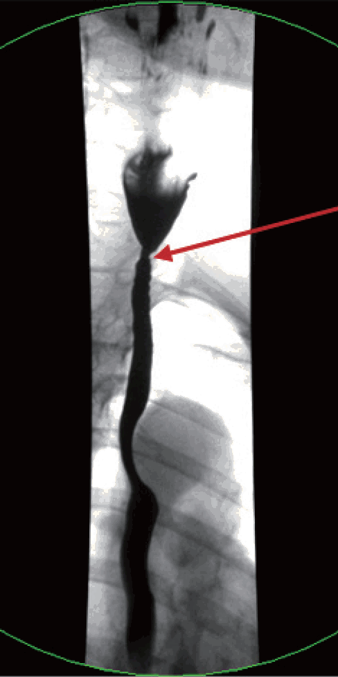
The esophageal stricture was located 18 cm from the incisor.

Endoscopy reveals a severe stricture in the cervical esophagus.
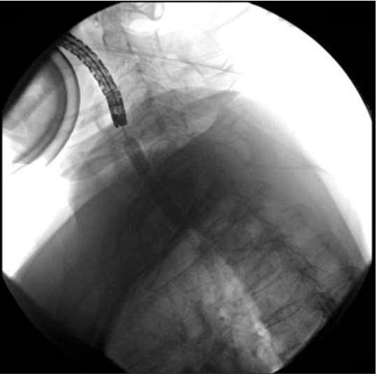
Fluoroscopic imaging of an unsuccessful attempt to use balloon dilation to treat the narrowed segment.
Incisional therapy using an insulated-tip electrosurgical knife.
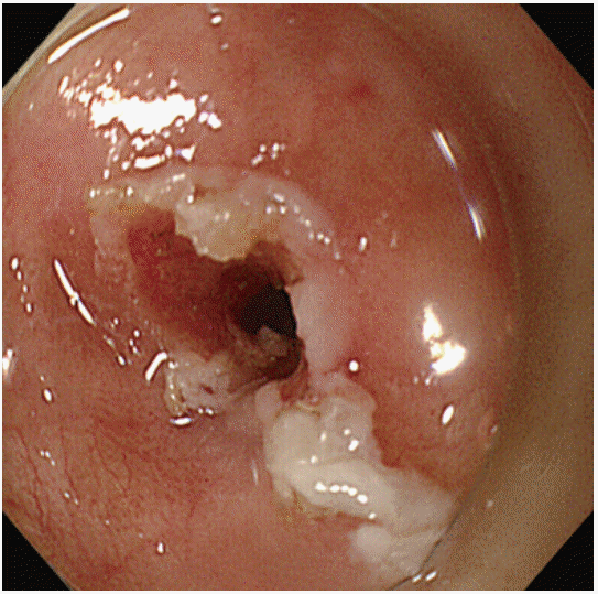
Endoscopy immediately after the incisional therapy reveals marked improvement at the stricture.
DISCUSSION
Approximately one-third of patients who experience a caustic esophageal injury subsequently develop an esophageal stricture and the resulting dysphagia can occur 2 weeks after the injury [8,9]. The cardinal feature of a caustic stricture is dysphagia because of esophageal narrowing or motility abnormalities, which are closely related to the scarred segment [10]. More than 80% of caustic strictures can be successfully treated with dilation [1,11], and bougie and balloon dilators are equally effective for treating benign lower esophageal strictures [2]. However, approximately one-third of these cases subsequently develop recurrent dysphagia within 1 year after dilation [3]. Consequently, patients with caustic strictures require a median of five sessions to achieve adequate dilation, compared to three sessions for patients with peptic strictures [1]. Unfortunately, a long-lasting response after dilation cannot be guaranteed for non-peptic strictures [5], and the treatment’s effect and re-treatment frequency often depends on the stricture’s etiology, length, and severity [5]. We classify esophageal strictures as simple or complex, and the complex strictures are more difficult to treat and tend to be refractory or recurrent, despite dilation therapy. Caustic strictures typically progress to the complex type [11,12], and strictures that are long (>2 cm), angulated, irregular, or severely narrowed tend to be refractory or recurrent, despite dilation therapy [13].
Incisional therapy has been reported as a treatment for anastomotic stricture after gastrointestinal surgery, peptic esophageal stricture, Schatzki’s ring, esophageal caustic stenosis, esophageal stricture after chemoradiotherapy or endoscopic submucosal dissection, and in combination with another treatment modality [14]. The success rate of incisional therapy is 80.6% for anastomotic strictures after gastrointestinal surgery [15], and incisional therapy was also successful in 16 of 20 peptic stricture cases (80%) [16]. Wills et al. [17] compared incision and bougie dilation for Schatzki’s rings, and reported more favorable outcomes for incisional therapy, which provided a longer symptom-free period and a greater reduction in dysphagia scores at 1 month. Canhoto et al. [6] described successful incisional therapy for a refractory esophageal caustic stricture in a 44-year-old man, who was free from dysphagia at the 1-year follow-up. Hagiwara et al. [18] evaluated the usefulness of incisional therapy combined with balloon dilation for cicatricial anastomotic strictures, and reported that five of the six patients exhibited both subjective and objective improvements for >4 weeks.In the present case, the patient’s odynophagia occurred 2 days after the treatment, and we believe that the odynophagia might be related to marked edema and narrowing caused by the electric currents from the incisional therapy, rather than the bougienage after the incision.
There is a report that local steroid injection therapy after endoscopic balloon dilation for benign esophageal stricture can decrease the recurrence of stricture [19]. Furthermore, Kim et al. [20] recently reported that oral steroid therapy after endoscopic submucosal dissection for an esophageal stricture can help prevent relapse after endoscopic balloon dilation. Therefore, in the present case, we used oral steroid therapy to help ameliorate the esophageal inflammatory response and prevent stricture recurrence.
In conclusion, we report a rare case of an intractable caustic esophageal stricture that was successfully managed with sequential treatment that comprised incision with an insulated-tip knife, balloon dilation, and an oral steroid. However, additional studies are needed to confirm the usefulness of this procedure for caustic esophageal strictures.
Notes
Conflicts of Interest: The authors have no financial conflicts of interest.