Benign bile duct strictures are commonly caused by stones, bile duct injury, sclerosing cholangitis, chronic pancreatitis, and congenital abnormalities. Bile duct changes caused by portal hypertension represent another group of uncommon causes of benign stricture.
The earliest evidence of portal biliopathy was described by Hunt in 1965 [1]. Portal cavernoma biliopathy (PCB) is a rare condition caused by cavernomatous transformation of an extra-hepatic portal vein thrombosis (EHPVT). It is rarely symptomatic; however, due to compression of the biliary tree, it can result in biliary obstruction, choledocholithiasis, jaundice, and cholangitis.
Endoscopic therapy for symptomatic PCB is a matter of discussion since this condition is rare [2].
We present a rare case of symptomatic PCB with a common bile duct (CBD) stricture, refractory to conventional endoscopic therapy in a patient unfit for surgery. Owing to the risk of repeated plastic stent exchange, we chose a more definite endoscopic approach with temporary placement of fully-covered self-expandable metal stent (fcSEMS). In this paper, we also report our successful results following application of fcSEMS.
An 82-year-old man presented to our hospital with fever, right-upper quadrant abdominal pain, and jaundice. He had undergone cholecystectomy at another hospital for acute cholecystitis in 2002, complicated with an EHPVT. In addition, he had a history of aortic stenosis, hypertension, and congestive heart failure.
His laboratory values were indicative of leukocytosis (16.700/uL) and elevated liver enzymes, with a total bilirubin of 8.9 mg/dL.
Abdominal ultrasound revealed dilated intrahepatic bile ducts (IHBD) and CBD, as well as an EHPVT.
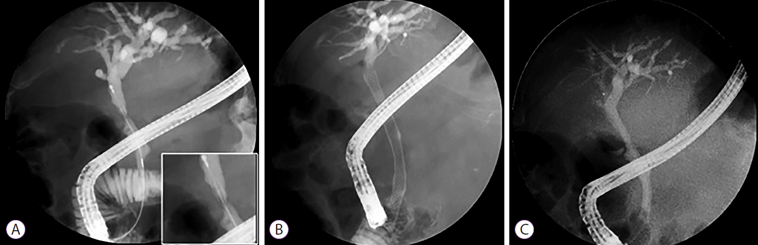
The patient was diagnosed with acute cholangitis and treated with antibiotics. Endoscopic retrograde cholangio-pancreatography (ERCP) showed asymmetric stenosis in the middle portion of the CBD (compatible with portal biliopathy type I2) and some small stones above it, with dilation of the proximal CBD and IHBD (Fig. 1A). After sphincterotomy and careful stone extraction with Dormiaтs basket, a 10-Fr plastic stent was placed. Balloon dilation was avoided due to hemobilia risk, which has been reported before [3,4].
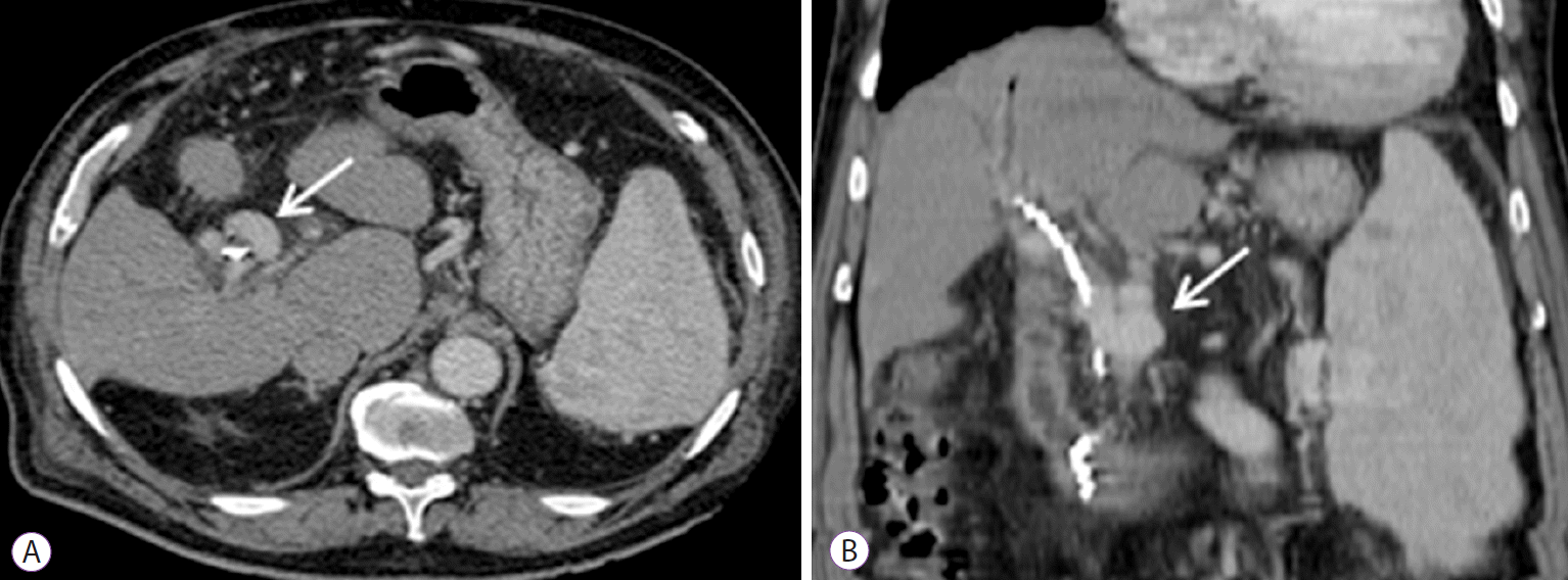
Abdominal computed tomography (CT) scan with contrast revealed a portal cavernoma compressing the middle segment of the CBD and causing the stricture seen during ERCP (Fig. 2).
The patient was not a candidate for portal decompression (transjugular portosystemic shunt was not feasible and he was unfit for surgery due to his advanced age and comorbidities), so progressive biliary plastic stenting was performed every 3 months. However, after one year, there was no improvement in the CBD stenosis and the patient still had elevated liver enzymes and IHBD dilation.
Following failure of progressive plastic stenting, temporary placement of a 80У10-mm fcSEMS (WallflexТЎ; Boston Scientific, Marlborough, MA, USA) was planned. The stent was deployed (Fig. 1B) and removed 6 months later (thereafter) without complications and revealing improvement of the stricture (Fig. 1C).
Liver enzymes became normal, the patient remained asymptomatic, and there was no need for additional endoscopic therapy in the last 12 months.
Currently, data on the endoscopic treatment of portal biliopathy strictures are insufficient [5]. As for endoscopic therapy, the risk of hemobilia has been studied as a major concern during endoscopic management of PCB. In particular, intra-choledochal varices, masquerading as filling defects, may be the source of bleeding during calculi extraction. Moreover, manipulation of the bile duct also carries a significant risk of complications. Hemobilia has also been reported for other interventions [6] such as exchange of plastic stents, pneumatic balloon dilatation of stricture, and balloon-occluding cholangiogram during endoscopic management of PCB.
Therefore, surgery is advised for symptomatic cases of PCB, whenever possible.
When surgery is not feasible or the patient is not fit for surgery (which was the case of our patient due to advanced age and comorbidities), plastic stenting is advised. However, it is also important to remember that repeated plastic stenting carries a possible risk of complications and, in refractory strictures, infinite exchanging of stents should ideally be avoided.
There are no guidelines regarding the use of SEMS in symptomatic PCB mainly due to very limited experience [5]. Moreover, cases managed with a successful and uncomplicated temporary placement of fcSEMS have been rarely reported [7,8]. Apart from possible migration, as with other strictures, excessive bleeding has been reported during removal of the metallic stent [9]. However, in this report, a fcSEMS was the first treatment approach, which is not routinely recommended.
According to the results obtained in our case, we believe temporary placement of fcSEMS might be a good option when surgery is contraindicated, progressive plastic stenting fails, and no episodes of hemobilia have occurred during previous endotherapy. It is reasonable to think that if bleeding has not occurred during plastic stent exchanges, the risk for it to occur during removal of a metal stent is smaller. However, more data are needed regarding this PCB treatment approach.