Introduction
Foreign body (FB) ingestion in children is very common, and most events occur in children between 6 months and 3 years of age. Notably, 80%ŌĆō90% of FBs in the gastrointestinal (GI) tract are passed spontaneously without complications, 10%ŌĆō20% are removed endoscopically, and 1% require open surgery secondary to complications [1]. Thus, FB ingestion presents a significant clinical difficulty in pediatric gastroenterological practice. In 2000, the American Association of Poison Control Centers documented that 75% of the >116,000 FB ingestions reported occurred in children aged Ōēż5 years [2].
Most ingested FBs are passed spontaneously through the GI tract without complications although endoscopic or surgical removal is required in a few children. However, optimal indications and/or timing of these procedures to be performed in children remain controversial. Fortunately, >90% of esophageal FBs are removed spontaneously without complications; however, a few cannot easily pass through the pylorus, stomach, duodenum, ileocecal valve, MeckelŌĆÖs diverticulum, and/or anus [3] and therefore, 10% of ingested FBs may remain in the GI tract [4,5].
Parameters that need to be considered regarding the timing of endoscopy in children with ingested FBs are the childrenŌĆÖs age or body weight, the clinical presentation, time since the last meal, time lapse since ingestion, type, as well as the size and the shape of the FB, and its present location in the GI tract [6].
Recently, owing to developments in and greater awareness of the usefulness of upper GI endoscopy in children, endoscopic removal of FBs is commonly considered an option in addition to waiting for spontaneous passage.
If endoscopic removal of the FB is not an emergency, or if it is not an absolute indication, the risk-benefit ratio ought to be considered in terms of assessing the complications expected to occur owing to the FB itself and those secondary to the procedure of FB removal.
Children characteristics such as age and weight vary, as do the type and size of the ingested FBs. Additionally, endoscopic removal of FBs is more difficult in young children than in adults. Therefore, it is difficult to determine an appropriate timing for endoscopic removal of FBs.
In this article, the author has reviewed the types and characteristics of FBs in the pediatric GI tract, and the indications and precautions pertaining to endoscopic removal of FBs.
Location
Esophagus
Unlike adults, young children accidentally swallow FBs. Esophageal FBs should be suspected in children who present with a sore throat, or difficulty swallowing saliva or food without an obvious reason.
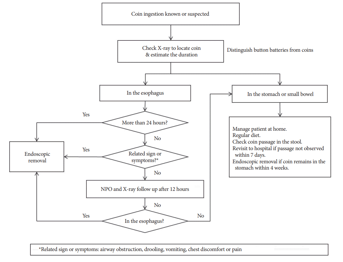
If an esophageal FB is not passed spontaneously within 24 hours, it must be removed considering the possibility of an anatomical anomaly or esophageal perforation [7,8].
Recently, the North American Society for Pediatric Gastroenterology, Hepatology, and Nutrition (NASPGHAN) Endoscopy Committee revised the recommendations pertaining to the timing of endoscopic intervention. The presence of esophageal button batteries mandates emergency removal within 2 hours regardless of the presence of symptoms [9].
Coins, magnets, sharp FBs, or food impaction in the esophagus all mandate removal within 2 hours if the childrenŌĆÖs secretions cannot be controlled. In asymptomatic children, they may be removed within 24 hours.
Long FBs lodged in the esophagus mandate removal within 24 hours regardless of the presence of symptoms.
Stomach
The NASPGHAN Endoscopy Committee recommends button battery removal within 2 hours in a symptomatic children regardless of size [9]. A button battery Ōēź20 mm located in the stomach of an asymptomatic children aged <5 years should be removed within 24 to 48 hours. If serial X-rays do not show progressive movement of an ingested FB in asymptomatic children, it can be observed for 24 hours. Magnets retained in the stomach in symptomatic children require removal within 2 hours. In asymptomatic children, they should be removed within 24 hours. Coins in the stomach of symptomatic children should be removed within 24 hours. In asymptomatic children, these can be observed for 24 hours. Long or large FBs in the stomach necessitate removal within 24 hours.
Small bowel
Most FBs in the small bowel are passed spontaneously without complications. Therefore, physicians should reassure the children and/or caregivers and advise them to check the childrenŌĆÖs stool for the FB. If the FB is not eliminated even after a week, children need to visit the hospital and obtain an X-ray to identify the accurate location of the FB.
Children should be strictly advised of the need to visit the hospital earlier if they develop signs of perforation or obstruction of the intestine, such as vomiting, severe abdominal pain, fever, or intestinal bleeding.
TYPES of foreign bodies
Coins
Coins are the most commonly ingested FB in children. Over 250,000 coin ingestions in children have been reported in the United States [10]. Factors influencing the spontaneous passage of a coin are its location in the esophagus, age of the child, and the size of the coin. Usually, the rate of spontaneous passage of swallowed coins in children is approximately 30% [11]. Thus, children presenting with an ingested coin without complications (a single coin lodged for <24 hours, without any history of esophageal disease or surgery, and no respiratory symptoms) can be observed over 12ŌĆō24 hours before performing an invasive procedure (endoscopic or surgical removal). Conners et al. suggested that coins lodged in the upper and mid esophagus require endoscopic removal, although 60% of coins lodged in the lower esophagus have been observed to pass spontaneously [12]. Once coins are observed to successfully pass through the esophagus, they are likely to progress and pass spontaneously [8,13,14]. Coins measuring >23.5 mm in size are more likely to become impacted, particularly in children aged <5 years. Coins measuring >25 mm in diameter are unlikely to pass through the pylorus, particularly in younger children even though they might have successfully passed through the esophagus [15]. Children in whom coin ingestion is observed or suspected need to undergo an X-ray to confirm the presence, size, and location of the coin, and the examination should be performed with close attention to distinguish the coin from a button battery, which shows the characteristic double halo sign (Fig. 1). Esophageal coins must be removed within 24 hours to reduce the incidence of complications. Symptomatic children presenting with difficulty swallowing saliva or respiratory difficulties warrant emergency endoscopic removal. After removal of esophageal coins, careful endoscopic examination of the esophageal mucosa is required to assess any evidence of significant injury.
Ingested coins present in the stomach can be observed in asymptomatic children in whom stool should be monitored for the passage of the coin, and serial X-rays should be obtained every 1 or 2 weeks until passage of the coin has been confirmed. If the coin is observed to remain in the stomach even after 2ŌĆō4 weeks, elective endoscopic removal can be considered. If the coin is located within the small bowel but the children are asymptomatic, clinical observation is indicated. However, in children presenting with symptoms of bowel obstruction or perforation, surgical removal needs to be considered (Fig. 2).
Button batteries
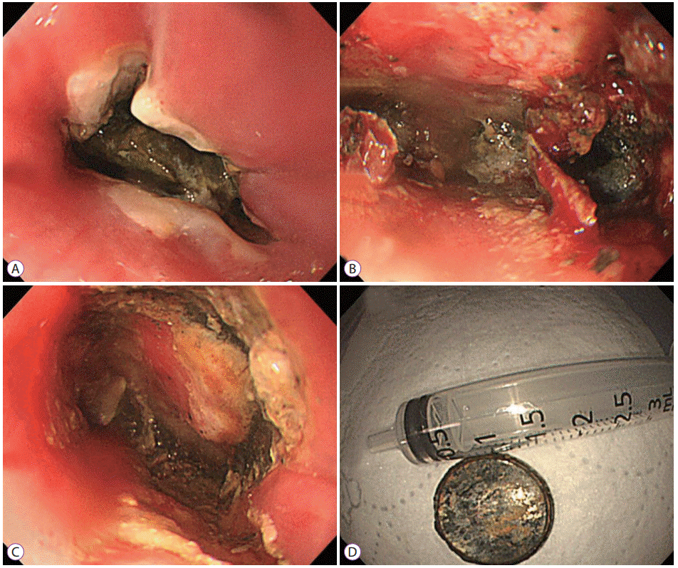
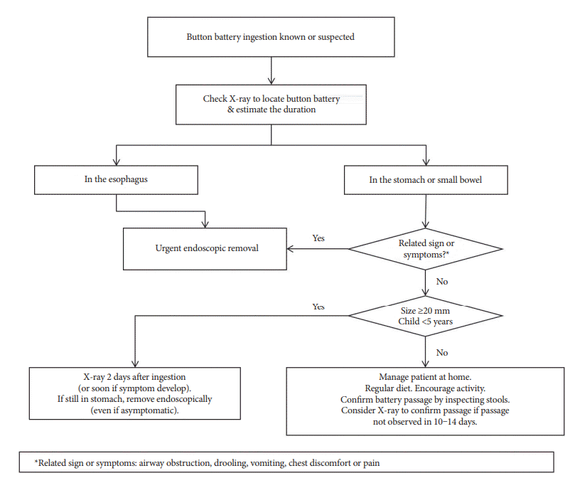
The frequency of button battery ingestion has been increasing owing to the widespread use of such batteries as power sources in electronic devices [16]. Button batteries resemble coins in size and shape; thus, because these two FBs are often indistinguishable, a careful X-ray examination is important to avoid a delay in diagnosis. Button batteries can cause severe damage secondary to local hydrolysis and the action of hydroxide on the mucosa, caustic injury secondary to a high pH, and minor electrical burns secondary to lithium. Button batteries impacted within the esophagus can cause burns within 4 hours. Usually, small button batteries (diameter Ōēż20 mm) do not cause serious complications that are observed in association with larger button batteries (diameter Ōēź20 mm) [17]. A study has shown that all 7 children who ingested button batteries <15 mm in size were asymptomatic without any complications, whereas all 5 children who swallowed batteries >15 mm in size showed moderate (n=3) to severe (n=2) complications [18]. The author described a 13-month-old infant who had ingested a 15-mm sized button battery 24 hours prior to presentation. He presented to the emergency room with vomiting and poor oral intake over a day prior to presentation. Unfortunately, nobody was aware that he had ingested the FB; however, an X-ray showed a round metal FB with a halo sign in his upper esophagus. An emergency endoscopic examination revealed a button battery that had caused an ulcer and corrosion of the esophageal mucosa (Fig. 3). Young children presenting with uncertain/undetermined evidence of ingested FBs need special attention.
The NASPGHAN Endoscopy Committee recommends removal of esophageal button batteries within 2 hours [9]. However, endoscopic removal of button batteries from the stomach remains a controversial issue. A large cohort study has shown that no previous reports have described significant gastric injury from button batteries [17]. Thus, the NASPGHAN Endoscopy Committee recommends observation of asymptomatic children (aged Ōēź5 years) who present with a short duration of ingestion (<2 hours) of a small-sized battery (<20 mm). Large batteries (>20 mm) remaining after 48 hours require removal (Fig. 4) [18].
Magnets
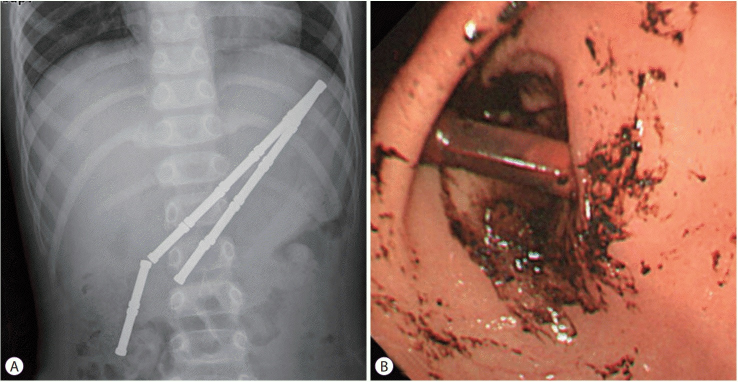
Recently, the frequency of magnet ingestion has increased in children. If a single magnet is ingested, it can be expected to be passed spontaneously if the magnet is not too large. However, if multiple magnets or a single magnet with a metallic FB has been ingested, the contact between these ingested magnets or the magnet and the metallic FB and the mucosal surfaces of different body parts can cause mucosal pressure necrosis, as well as intestinal obstruction, fistula, and/or perforation; therefore, surgical removal is needed in such cases [19-21].
If magnet ingestion is detected on an X-ray, the physician must confirm whether the ingested FBs are single or multiple magnets or magnets with a metallic FB. Occasionally, two or more magnets may be attached to each other and may appear like one piece, and misdiagnosis of multiple magnets as solitary magnet ingestion can lead to delayed institution of treatment and cause significant complications. Given this risk, if multiple magnets or a single magnet with a metallic FB are located within the esophagus or the stomach, these FBs must be endoscopically removed even in asymptomatic children (Fig. 5).
If multiple magnets or a single magnet with a metallic FB are located in sites beyond the stomach, symptomatic children need to consult a pediatric surgeon to plan surgery and asymptomatic children may be closely followed using serial X-rays to monitor progression of the FBs.
Recently newer and smaller neodymium magnets that are at least 5 to 10 times stronger than traditional magnets are available as adult toys and can attract each other with powerful forces [22]. A neodymium magnet appears like a ball-bearing on an X-ray, and clinicians should be careful to not misdiagnose it as a metal ball.
Sharp or pointed foreign bodies
Ingestion of sharp or pointed FBs in children is known to be associated with high morbidity and mortality, and delayed diagnosis and management increases the risk of serious complications.
Sharp or pointed FBs such as safety pins, nails, hair-pins, screws, pine needles, thumbtacks, or dental prostheses can cause serious complications such as esophageal ulceration and/or perforation, trachea-fistula, and/or abscess formation, peritonitis, an aorto-esophageal fistula, and even death [23-26]. Usually, intestinal FBs are known to cause perforation in <1% of patients; however, sharp or pointed FBs can cause perforation in 15%ŌĆō35% of patients. Therefore, it is preferable to remove FBs from the esophagus or stomach whenever possible. Notably, in recent times, early diagnosis and prompt endoscopic removal have reduced the incidence of adverse events related to the ingestion of sharp or pointed FBs [27]. Early diagnosis requires accurate information regarding the childrenŌĆÖs history or a high index of clinical suspicion for the ingestion of a sharp FB and an urgent X-ray examination. Radiolucent FBs such as plastic, glass, fish bones or wood cannot be identified using X-ray examination. Thus, in children with suspected ingestion of sharp FBs, even if an X-ray does not reveal a FB, an emergency endoscopy is recommended. A sharp FB present in the esophagus constitutes a medical emergency because of the high risk of perforation and migration and warrants emergency removal even if the children have not been maintained on a nil per os status. Overtubes may be utilized during endoscopic variceal band ligation when removing sharp FBs in adults, although their use is difficult in children because of a large diameter. Removal of sharp FBs using an endoscopic cap can prevent esophageal injury in children. If the sharp end of the FB is observed to be facing the proximal site, it may be safest to push the FB into the stomach and rotate its sharp end toward the distal site before removal. Sharp or pointed FBs, long objects (>4ŌĆō5 cm in infants and young children, those >6ŌĆō10 cm in older children), or large and wide objects (>2 cm in diameter in infants and young children, >2.5 cm in diameter in older children) that are located in the stomach, warrant endoscopic removal [1]. If a sharp FB has passed into the small bowel (distal to the ligament of Treitz), surgical removal can be considered in symptomatic children. In asymptomatic patients, close clinical follow-up with serial X-rays obtained after admitting the patient are recommended. The mean GI transit time for FBs in children is approximately 3.6 days [28]. Therefore, if the FB does not show the expected passage after 4 days, a bowel perforation or a congenital anomaly is suspected, and surgical removal of the FB needs to be considered [1,29,30].
Large or long foreign bodies
Ingestion of large or long FBs is an issue of special concern. These FBs must be removed within 24 hours because long (those >6 cm in length) or large FBs are unlikely to pass through the duodenum and the ileocecal valve [31].
Sharp or pointed objects, long objects (>4ŌĆō5 cm in infants and young children, >6ŌĆō10 cm in older children), or large and wide objects (>2 cm in diameter in infants and young children, or >2.5 cm in diameter in older children) located in the stomach warrant endoscopic removal [1].
Fish bones
Fish bones comprise the most common food-related FB ingested by children. Both Korea and China, which show a high intake of fish demonstrate a higher incidence of fish bone ingestion than that in other countries [32].
Children usually show fish bone impaction in the palatine tonsils, tongue base, vallecula and pyriform sinus because the laryngopharynx is narrower and the tonsils are larger in children than in adults. A Korean study has reported that ingested fish bones in children were most commonly detected in the pharynx (57.7%) [6]. In fact, fish bone impaction is rare in the esophagus below the pharynx. However, fish bones lodged in the esophagus can cause mucosal ulceration or a topical inflammatory reaction leading to esophageal stenosis, perforation, a deep neck abscess, mediastinitis, a lung abscess, or even aortic fistulae. Therefore, prompt and accurate diagnosis and treatment are required.
Conclusions
Children with upper GI FB ingestion can be effectively treated by an experienced endoscopist with safe and uncomplicated removal of such FBs using pediatric and appropriate ancillary endoscopic equipment.
However, it is necessary to carefully consider the type of FB ingested, the childrenŌĆÖs age, expected complications, and emergency situations. It is also important to establish effective coordination between a medical delivery system as well as medical personnel and equipment.