A Rare Duodenal Subepithelial Tumor: Duodenal Schwannoma
Article information
Abstract
Schwannomas are uncommon neoplasms that arise from Schwann cells of the neural sheath. Gastrointestinal schwannomas are rare among mesenchymal tumors of the gastrointestinal tract, and only a few cases have been reported to date. Duodenal schwannomas are usually discovered incidentally and achieving a preoperative diagnosis is difficult. Schwannomas can be distinguished from other subepithelial tumors on endoscopic ultrasonography; however, any typical endosonographic features of duodenal schwannomas have not been reported due to the rarity of these tumors. Immunohistochemistry is essential to distinguish schwannomas from gastrointestinal stromal tumors and leiomyomas. We report a case of duodenal schwannoma found incidentally during a health check-up endoscopy. On endoscopic ultrasonography, this tumor was suspected as a gastrointestinal stromal tumor; therefore, the patient underwent laparoscopic wedge resection of the tumor. Histopathology and immunohistochemistry confirmed that the duodenal lesion was a benign schwannoma.
Introduction
Schwannomas, also known as neurilemmomas, are benign tumors derived from Schwann cells of the neural sheath. These tumors are rare among mesenchymal tumors of the gastrointestinal (GI) tract and the incidence is 2% to 6% in all subepithelial tumors of the GI tract [1]. GI schwannomas occur most commonly in the stomach, followed by the colon and rectum [2]. Meanwhile, schwannomas of the small intestine have rarely been reported [3]. The clinical course of GI schwannomas is not specific and their preoperative diagnosis is difficult. Schwannomas can be distinguished from other subepithelial tumors on endoscopic ultrasonography (EUS) and immunohistochemistry. A positive reaction for S-100 protein turns out to be supportive for the diagnosis of schwannomas [3]. Herein, we report the case of a patient with schwannoma of the duodenum confirmed by surgical resection.
CASE REPORT
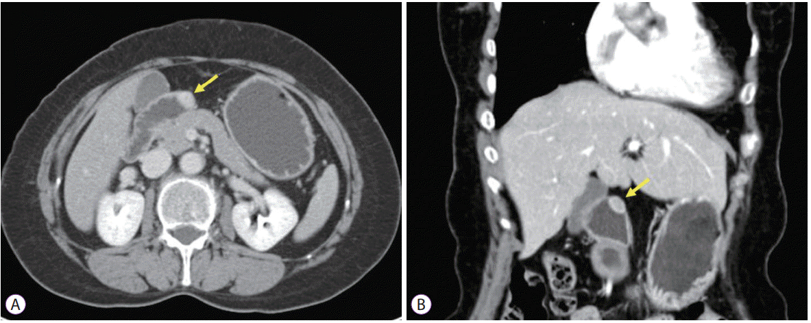
A 54-year-old woman was transferred to our hospital for further evaluation and management for a subepithelial tumor on the duodenum. Any specific symptoms and signs were not seen on physical examination of the patient. There was no history of upper GI bleeding, or past medical or surgical interventions. The vital signs and laboratory findings were within the normal range. On upper GI endoscopy, a subepithelial mass was seen in the duodenal bulb, and its surface was covered by normal duodenal mucosa (Fig. 1A). On EUS, the mass was heterogeneously hypoechoic with marginal haloes that had originated from the muscularis propria layer (Fig. 1B). This lesion was suspected as a GI stromal tumor (GIST). An abdominopelvic computed tomography (CT) showed an ovoid homogenously-enhancing mass measuring approximately 1.5×1.0 cm in the anterior wall of duodenal bulb (Fig. 2). Neither obvious metastasis to surrounding organs nor intra-abdominal lymphadenopathy was observed. Since this lesion was strongly suspected as a GIST, surgical intervention was planned. The patient underwent laparoscopic wedge resection of the tumor. Grossly, the tumor was an intramural, solid mass and measured 2.0×1.7 cm in diameter. On cross section, the tumor was yellowish-to-white in color (Fig. 3A). Microscopically, the tumor grew intramurally without capsule, which was located in the muscularis propria. On hematoxylin-eosin staining, the tumor was composed of spindle-shaped cells arranged in a palisading nuclei pattern forming cellular and hypocellular areas (Fig. 3B). Mild nuclear atypia was seen, but the mitotic activity was less than 5/50 high-power fields. The tumor cells were strongly positive for S-100 protein, but negative for smooth muscle actin, CD34, and c-kit (Fig. 3C-F). Finally, the subepithelial tumor was diagnosed as a schwannoma of the duodenum. The patient’s postoperative course was uneventful and she was discharged on the fifth postoperative day. At 8 years follow-up, currently, the patient remains free from the disease.

(A) Upper endoscopy reveals a subepithelial mass covered by the normal mucosa on the duodenal bulb. (B) On endoscopic ultrasonography, the mass is a 1.5×0.7 cm sized heterogeneously hypoechoic mass with marginal haloes originating from the muscularis propria layer.

Abdominopelvic computed tomography scans show a 1.5×1.0 cm sized homogenously-enhancing mass in the anterior wall of the duodenal bulb (arrow). (A) Axial section view. (B) Coronal section view.

(A) The resected tumor is an intramural, solid, yellowish-to-white mass on cross section. (B) Microscopically, the mass is composed of spindle cells with wavy nuclei. Mild nuclear atypia are seen, but the mitosis count is less than 5/50 high-power fields (hematoxylin and eosin stain, ×200). Immunohistochemically, the tumor cells are strongly positive for S-100 protein, ×400 (C), but negative for smooth muscle actin, ×400 (D), CD34, ×400 (E), and c-kit, ×400 (F).
DISCUSSION
Schwannomas are uncommon neoplasms arising from Schwann cells, which cover the peripheral nerves, and are difficult to distinguish from other mesenchymal tumors. The most common GI site of schwannomas is the stomach, and the duodenum is extremely rare [4]. Duodenal schwannomas are mostly located in the second or third portion of the duodenum [5]. Nilsson et al. reported that, of 43 small intestinal schwannomas, eight were located in the duodenum [6]. There was no difference in the incidence between men and women, and most occurred in the fifth to sixth decades of life [3]. Our patient was in the sixth decade and the tumor was located in the duodenal bulb, which differs from that which has been reported in the literature. Duodenal schwannomas are usually asymptomatic and can be discovered incidentally. If symptoms are present, the most common symptom is intestinal hemorrhage and abdominal discomfort [5]. Duodenal schwannomas are usually benign, and malignant transformation is very rare. There are no histologic criteria to determine the grade of schwannoma and the metastatic potential is not yet known. However, malignant changes are reported in peripheral schwannomas [7]. Therefore, careful observation after surgery is sometimes needed.
For diagnosis of schwannomas, endoscopy, EUS, abdominal CT, and abdominal magnetic resonance imaging (MRI) are useful to determine localization, relationship of surrounding organs, tumor multiplicity, or metastasis. Since the growth of the schwannomas is subepithelial, endoscopic evaluation is difficult to distinguish schwannomas from other mesenchymal tumors, such as GISTs, leiomyomas, or leiomyosarcomas. Furthermore, endoscopic biopsy is not suitable for definitive diagnosis because the schwannoma is a subepithelial tumor and mucosal abnormalities are rarely seen [8]. EUS and EUS-guided fine-needle biopsy (EUS-FNB) are very valuable techniques for performing directed biopsies when necessary [9]. However, any typical EUS features of duodenal schwannomas have not been reported due to the rarity of these tumors. In the case of gastric schwannomas, several typical EUS features, such as heterogeneously or homogenously hypoechoic lesions with marginal haloes, and few internal echogenic foci are reported [10-12]. CT is the most common method for evaluating neurogenic tumors and is useful for distinguishing benign and malignant neoplasms and predicting preoperative stage. On CT, the schwannomas are seen as homogeneously enhancing masses with a round or oval shape, which project into the lumen of the small intestine [13]. In MRI, GI schwannomas are sharply demarcated and strongly enhancing tumors, with low to medium signal intensity on T1-weighted images and high signal intensity on T2-weighted images [14]. However, schwannomas cannot be distinguished from malignant neurogenic tumors by radiologic images alone. Therefore, histological examination is essential for definitive diagnosis. Schwannomas are encased by the intact mucosa and usually involve the submucosa and muscularis propria. These tumors are composed of spindle-shaped nuclei with high and low cellularity regions called Antoni A and Antoni B areas. Conventional hematoxylin and eosin staining dose not distinguish neurogenic and myogenic tumors. Immunohistochemistry is essential to distinguishing between schwannomas and GISTs or leiomyomas. Cells of schwannoma are 100% immunoreactive with S-100 protein. GISTs are usually positive for c-kit and CD34, but negative for S-100 protein, whereas leiomyomas are positive for smooth muscle actin and desmin, and negative for S-100 protein [1]. The treatment of choice is complete surgical resection, with an approach that depends on tumor size, localization, and histological features. The optimal treatment for a malignant schwannoma has not been fully established [3]. The role of chemotherapy and radiotherapy remains unclear. Incomplete resection can be associated with local recurrence. Thus, surgical margins have been regarded as the most important prognostic factor.
In conclusion, we report a very rare case of schwannoma that arises in the duodenal bulb. Since schwannomas are difficult to distinguish from other mesenchymal tumors, diagnosis and treatment may be delayed. A definitive diagnosis is difficult before surgical resection. Imaging studies with abdominal CT, EUS and MRI and recently EUS-FNB can increase the diagnosis rate before operation.
Notes
Conflicts of Interest:The author has no financial conflicts of interest.
