Skip Navigation
Skip to contents

Clin Endosc : Clinical Endoscopy

OPEN ACCESS

Articles

Page Path
HOME > Clin Endosc > Volume 53(4); 2020 > Article
CME for
KSGE members
Boost Your Learning With Quiz Rare Cause of a Colonic Laterally Spreading Tumor
Sung Min Lee1orcid, Dong Hae Chung2orcid, Kwang An Kwon1,orcid
Clinical Endoscopy 2020;53(4):499-501.
DOI: https://doi.org/10.5946/ce.2020.197
Published online: July 30, 2020

1Department of Gastroenterology, Gachon University Gil Medical Center, Gachon University College of Medicine, Incheon, Korea

2Department of Pathology, Gachon University Gil Medical Center, Gachon University College of Medicine, Incheon, Korea

Correspondence: Kwang An Kwon Department of Gastroenterology,Gachon University Gil Medical Center, Gachon University College of Medicine, 21 Namdong-daero 774 beon-gil, Namdong-gu, Incheon 21565, Korea Tel: +82-32-460-3778, Fax: +82-32-460-3408, E-mail: toptom@gilhospital.com
• Received: July 27, 2020   • Revised: July 28, 2020   • Accepted: July 28, 2020

Copyright © 2020 Korean Society of Gastrointestinal Endoscopy

This is an Open Access article distributed under the terms of the Creative Commons Attribution Non-Commercial License (http://creativecommons.org/licenses/by-nc/3.0/) which permits unrestricted non-commercial use, distribution, and reproduction in any medium, provided the original work is properly cited.

  • 4,155 Views
  • 88 Download
prev
A laterally spreading tumor was found during a screening colonoscopy in a 71-year-old woman. The lesion was flat and irregular shaped, 20 mm × 30 mm in size, and located 10 cm above the anal verge. There was no ulcer or erosion on the lesion. For histologic diagnosis and therapeutic purpose, a piecemeal endoscopic mucosal resection was performed. The endoscopic and histologic images are shown in Fig. 1.
What is the most likely diagnosis?
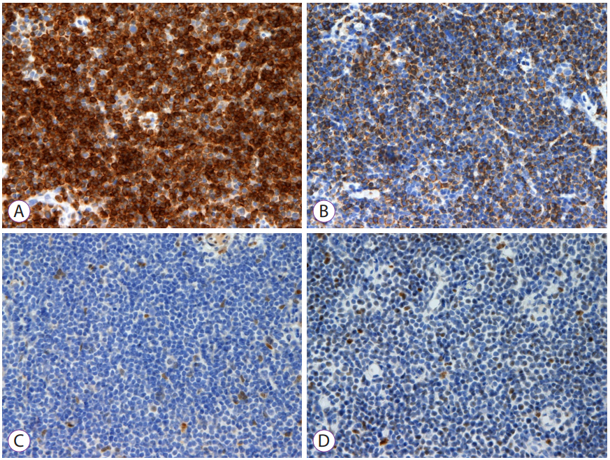
The immunohistochemistry results are described in Fig. 2.
Histopathological examination of the specimen revealed that small-sized lymphocytes infiltrated into the mucosal and submucosal layers. The results of the immunohistochemical stain were as follows: CD5(–), CD20(+), cyclin D1(–), Bcl-2(+), and Bcl-6(–). An appropriate diagnosis is colonic mucosa-associated lymphoid tissue (MALT) lymphoma because tumor cells were positive for CD20 and Bcl-2, whereas negative for cyclin D1 and Bcl-6. For staging, abdominopelvic computed tomography, chest computed tomography, and positron emission tomography-computed tomography were performed. No metastatic lesions were found. The patient is currently undergoing follow-up in outpatient clinic without progression of the disease.
MALT lymphoma most often occurs in the stomach, in addition to other organ systems such as the thyroid and salivary glands, lungs, and breasts [1]. Gastrointestinal MALT lymphomas can occur in the small and large intestines in addition to the stomach. About 80% of gastric MALT lymphomas are thought to be associated with Helicobacter pylori infections. Chronic inflammation of epithelial cells due to a H. pylori infection causes proliferation of T cells in the mucous membrane and proliferation of B cells. Among them, the proliferation of B cells with genetic mutations results in MALT lymphoma. In addition, MALT lymphomas have been reported to be related to Borrelia burgdorferi, Chlamydia jejuni, and hepatitis C virus infections and some autoimmune diseases [2].
Systemic symptoms, such as B symptoms, are rare because colonic MALT lymphomas have a slow-growing nature and remain in the local area. Colonic MALT lymphoma is most commonly found around the ileocecal valve area, often in order of occurrence starting from the rectum, ascending colon, transverse colon, sigmoid colon, and descending colon.
Endoscopic findings of colonic MALT lymphomas are predominantly in the form of a single polyp and sessile elevated mucosal lesion [3]. The endoscopic findings of gastric MALT lymphomas can be diverse, such as superficial ulcers, irregular mucosa, atrophic mucosa, bumps, and erosion. Gastrointestinal MALT lymphoma is diagnosed by an endoscopic biopsy and immunohistochemical staining for suspected lesions. Gastrointestinal MALT lymphoma can be diagnosed by lymphoepithelial lesions due to infiltration of atypical lymphocytes, centrocyte-like cells, and plasma cell differentiation as well as by cells that are positive for CD19, CD20, CD22, and bcl-2 and negative for CD3, CD5, CD10, and bcl-1/cyclin D1 [4]. The MALT lymphoma shows positive for B cell markers (CD19, CD20, and CD22), and low-grade B-cell non-Hodgkin lymphoma among the extranodal marginal zone lymphomas has similar immunohistochemical staining results of being negative for CD5, CD10, and CD23.
In addition, endoscopic ultrasound, computed tomography, and bone marrow biopsy are performed for staging.
Treatment of gastrointestinal MALT lymphoma depends on the stage. In cases of gastric MALT lymphoma, H. pylori eradication is performed first at an early stage, and radiotherapy and chemotherapy are administered in cases of initial treatment failure. The effectiveness of H. pylori eradication in MALT lymphomas occurring in areas other than the stomach varies. For Colorectal MALT lymphomas, there are yet no clear treatment guidelines and various treatments such as endoscopic resection, surgery, combined chemotherapy, local radiation therapy, and H. pylori eradication are used [5,6].
Fig. 1.
Colonoscopy and microscopy findings. (A-C) Colonoscopy revealed a flat, irregularly marginated, and superficially elevated lesion at the mid-rectum for which a piecemeal endoscopic mucosal resection was performed; (D) Microscopy revealed dense lymphoid infiltration involving the mucosa and submucosa (hematoxylin and eosin, ×40).
ce-2020-197f1.jpg
Fig. 2.
Immunohistochemistry results. (A) Positive for CD20; (B) Positive for Bcl-2; (C) Negative for cyclin D1; (D) Negative for Bcl-6.
ce-2020-197f2.jpg
  • 1. Khalil MO, Morton LM, Devesa SS, et al. Incidence of marginal zone lymphoma in the United States, 2001-2009 with a focus on primary anatomic site. Br J Haematol 2014;165:67–77.ArticlePubMedPMC
  • 2. Lecuit M, Abachin E, Martin A, et al. Immunoproliferative small intestinal disease associated with Campylobacter jejuni. N Engl J Med 2004;350:239–248.ArticlePubMed
  • 3. Rhee JC, Lee HY, Rhee PL, et al. [Esophagus, stomach & intestine; endoscopic findings of gastric mucosa: associated lymphoid tissue (MALT) lymphoma]. Korean J Gastrointest Endosc 1997;17:125–132.
  • 4. Cavalli F, Isaacson PG, Gascoyne RD, Zucca E. MALT lymphomas. Hematology Am Soc Hematol Educ Program 2001;241–258.ArticlePubMedPDF
  • 5. Chung CH, Kim HG, Park WS, et al. [A case of mucosa-associated lymphoid tissue lymphoma of colon as multiple large polypoid lesions]. Korean J Gastrointest Endosc 2001;23:122–126.
  • 6. Lee YG, Lee S, Han SW, Lee JS. A case of multiple mucosa-associated lymphoid tissue (MALT) lymphoma of the colon identified as simple mucosal discoloration. J Korean Med Sci 2005;20:325–328.ArticlePubMedPMC

Figure & Data

REFERENCES

    Citations

    Citations to this article as recorded by  

      • PubReader PubReader
      • ePub LinkePub Link
      • Cite
        CITE
        export Copy Download
        Close
        Download Citation
        Download a citation file in RIS format that can be imported by all major citation management software, including EndNote, ProCite, RefWorks, and Reference Manager.

        Format:
        • RIS — For EndNote, ProCite, RefWorks, and most other reference management software
        • BibTeX — For JabRef, BibDesk, and other BibTeX-specific software
        Include:
        • Citation for the content below
        Rare Cause of a Colonic Laterally Spreading Tumor
        Clin Endosc. 2020;53(4):499-501.   Published online July 30, 2020
        Close
      • XML DownloadXML Download
      Figure
      • 0
      • 1
      Rare Cause of a Colonic Laterally Spreading Tumor
      Image Image
      Fig. 1. Colonoscopy and microscopy findings. (A-C) Colonoscopy revealed a flat, irregularly marginated, and superficially elevated lesion at the mid-rectum for which a piecemeal endoscopic mucosal resection was performed; (D) Microscopy revealed dense lymphoid infiltration involving the mucosa and submucosa (hematoxylin and eosin, ×40).
      Fig. 2. Immunohistochemistry results. (A) Positive for CD20; (B) Positive for Bcl-2; (C) Negative for cyclin D1; (D) Negative for Bcl-6.
      Rare Cause of a Colonic Laterally Spreading Tumor

      Clin Endosc : Clinical Endoscopy Twitter Facebook
      Close layer
      TOP