Skip Navigation
Skip to contents

Clin Endosc : Clinical Endoscopy

OPEN ACCESS

Articles

Page Path
HOME > Clin Endosc > Volume 54(3); 2021 > Article
Original Article Efficacy of Underwater Endoscopic Mucosal Resection for Superficial Non-Ampullary Duodenal Epithelial Tumor
Masanori Furukawa1,orcid, Akira Mitoro2orcid, Takahiro Ozutumi2orcid, Yukihisa Fujinaga2orcid, Keisuke Nakanishi2orcid, Koh Kitagawa2orcid, Soichiro Saikawa2orcid, Sinya Sato2orcid, Yasuhiko Sawada2orcid, Hiroaki Takaya2orcid, Kosuke Kaji2orcid, Hideto Kawaratani2orcid, Tadashi Namisaki2orcid, Kei Moriya2orcid, Takemi Akahane2orcid, Junichi Yamao1orcid, Hitoshi Yoshiji2orcid
Clinical Endoscopy 2021;54(3):371-378.
DOI: https://doi.org/10.5946/ce.2020.147
Published online: February 18, 2021

1Division of Endoscopy, Nara Medical University Hospital, Nara, Japan

2Department of Gastroenterology, Nara Medical University, Nara, Japan

Correspondence: Masanori Furukawa Division of Endoscopy, Nara Medical University Hospital, Shijo-cho 840, Kashihara, Nara 634-8522, Japan Tel: +81-744-22-3051, Fax: +81-744-21-7152, E-mail: furukawa@naramed-u.ac.jp
• Received: June 3, 2020   • Revised: August 30, 2020   • Accepted: September 7, 2020

Copyright © 2021 Korean Society of Gastrointestinal Endoscopy

This is an Open Access article distributed under the terms of the Creative Commons Attribution Non-Commercial License (http://creativecommons.org/licenses/by-nc/3.0/) which permits unrestricted non-commercial use, distribution, and reproduction in any medium, provided the original work is properly cited.

  • 4,950 Views
  • 166 Download
  • 18 Web of Science
  • 18 Crossref
  • 19 Scopus
prev next
  • Background/Aims
    Endoscopic resection (ER) for superficial non-ampullary duodenal epithelial tumors (SNADETs) is challenging. Conventional endoscopic mucosal resection (CEMR) is also problematic due to the anatomical features of the duodenum. We compared the safety and efficacy of underwater endoscopic mucosal resection (UEMR) with those of CEMR through a retrospective analysis.
  • Methods
    Altogether, 44 consecutive patients with 46 SNADETs underwent ER (18 CEMR cases and 28 UEMR cases) between January 2016 and October 2019. We investigated the proportions of en bloc resection, R0 resection, complications, resection time, and total procedure time and compared the outcomes of patients from the CEMR group with those of patients from the UEMR group.
  • Results
    The median tumor size was 8.0 mm (range, 2.0–20.0 mm). The UEMR group showed a higher proportion of en bloc resection (96.4% vs. 72.2%, p<0.05) and significantly lower median resection time and total procedure time (4 min vs. 9.5 min, p<0.05 and 13 min vs. 19 min, p<0.05; respectively) than the CEMR group. No complications were observed. However, two patients treated with piecemeal resection in the CEMR group had residual tumors.
  • Conclusions
    UEMR is a feasible therapeutic option for SNADETs. It can be recommended as a standard treatment.
Until recently, superficial non-ampullary duodenal epithelial tumor (SNADET) was considered a very rare disease, with estimated prevalence rate of 0.01%–0.4% [1-4]. However, due to advances in endoscopic technology, detection rate of SNADET is increasing [5]. Non-ampullary duodenal adenomas are precancerous lesions and therefore, require early therapeutic intervention [6,7]. With minimal lymph node metastasis, less invasive endoscopic treatment is ideal for SNADETs [8-10]. However, standard endoscopic resection (ER) has not been established as a treatment for SNADETs due to a considerable rate of adverse events. Conventional endoscopic mucosal resection (CEMR) is the most common treatment for SNADETs. However, CEMR for SNADETs has a high recurrence rate (5%–37%), with constant rates of adverse events such as delayed bleeding (0%–15%), intraprocedural perforation (0%–2%), and delayed perforation (0%–4%) [10-14]. Endoscopic submucosal dissection (ESD) is widely accepted as a high-quality treatment for superficial neoplasias of the gastrointestinal tract regardless of the lesion size. Duodenal ESD has been associated with a greater number of adverse events such as perforation and bleeding [15-18]. Underwater endoscopic mucosal resection (UEMR) eliminates the need for submucosal injection. It has been attracting attention as an excellent endoscopic treatment for SNADETs with very few adverse events [19,20]. The present study aimed to investigate the preferred endoscopic treatment (UEMR or CEMR) for SNADET.
Study design
The present retrospective analysis of ER for duodenal tumors was conducted at the Department of Gastroenterology of Nara Medical University (Nara, Japan). The study protocol adhered to the ethical principles of the Declaration of Helsinki and was approved by the institutional review board of Nara Medical University Hospital (approval no. 2049). All authors had access to the study data and approved the final manuscript. This manuscript is presented according to the Strengthening the Reporting of Observational Studies in Epidemiology (STROBE) Statement.
Patients
Consecutive patients with duodenal tumors sized ≤20 mm who underwent ER between January 2016 and October 2019 were enrolled in this study. Patients with neuroendocrine tumors or ampullary duodenal tumors were excluded. Patients’ data and the details of the procedures were retrieved from the hospital database and from the patients’ charts. In our department, CEMR (the “injection and snaring method” or the “two-channel strip biopsy method”) was performed from January 2016 to January 2018 and UEMR was performed from February 2018 to October 2019. We evaluated patient characteristics, endoscopic findings, treatment outcomes including histological findings, and adverse events between patients who underwent CEMR and those who underwent UEMR.
Procedures
All ER procedures were performed by board certified endoscopists of the Japan Gastroenterological Endoscopy Society with sufficient endoscopic treatment experience. CEMR was performed using either the “injection and snaring method” or the “two-channel strip biopsy method”. The “injection and snaring method” was performed principally using a therapeutic endoscope with a water jet function (GIF-Q260J; Olympus, Tokyo, Japan) and the “two-channel strip biopsy method” was performed using a two-channel endoscope (2T240; Olympus). For all EMR cases, a transparent cap (model D-201-11804 or D-201-13404; Olympus) was attached to the tip of the endoscope. Hypertonic saline-epinephrine or 0.4% sodium hyaluronate (Mucoup; Boston Scientific Japan, Tokyo, Japan) was injected into the submucosal layer before the initiation of both methods. Mucosal resection was performed using an electrocautery snare (Captivator II; Boston Scientific, Marlborough, MA, USA) powered by an electrosurgical generator (VIO300D; Erbe Elektromedizin, Tubingen, Germany). During the “injection and snaring” process for CEMR, the lesion elevated with a submucosal injection was captured with an electrosurgical snare and resected with electrocautery (Endo Cut Q mode, effect 3, duration 2, and interval 4). During the “two-channel strip biopsy”, a grasping forceps was inserted into the right channel of a two-channel endoscope and passed through an electrosurgical snare inserted from the left channel. The lesion elevated with a submucosal injection was grasped and pulled up with the forceps. The lesion was then captured and resected using an electrosurgical snare with electrocautery, similar to the “injection and snaring method”. UEMR was performed using a therapeutic endoscope with a water jet function (GIFQ260J; Olympus). For all UEMR cases, a transparent cap (model D-201-11804; Olympus) was attached to the tip of the endoscope. After complete deflation of the gastric and the duodenal lumens, 0.9% saline solution was infused until the lesion was adequately visualized in the saline. Three electrosurgical snare sizes were used according to the lesion size. Once the lesion was trapped, it was transected with electrocautery (Endo Cut Q mode, effect 3, duration 2, and interval 4) using the same electrosurgical generator used for CEMR. After resection with both the methods, any active hemorrhage was coagulated with hemostatic forceps (Coagrasper, FD410LR; Olympus) using the soft coagulation mode (effect 5, 80 W). Subsequently, the mucosal defect was observed carefully and additional resection was performed using the electrosurgical snare if an endoscopically apparent or suspicious residual tumor was observed. The surgeon attempted to close the mucosal defects with endoclips (EZ clip; Olympus) as frequently as possible to prevent delayed adverse events. Patients fasted for 2 days including the day of the resection procedure. After confirming that there were no adverse events, patients were allowed to begin a liquid diet on postoperative day (POD) 3 and were usually discharged on POD 6. Patients were administered proton pump inhibitors (lansoplazole 30 mg/day or esomeprazole 20 mg/day) or a potassium-competitive acid blocker (vonoprazan 20 mg/day) for 6 weeks. Resected specimens were embedded in 10% formalin and fixed specimens were sectioned serially at 2 mm intervals. Two experienced pathologists assessed the specimens according to the revised Vienna classification [21]. All patients underwent endoscopy at our hospital to assess any residual tumors 2 months after the procedure.
Data analysis and outcomes
Collected data included patients’ age, sex, lesion characteristics (location, size, macroscopic type), resection time, total procedure time, en bloc resection rate, R0 resection rate, pathological diagnosis, adverse events, and recurrence. The resection time was measured from submucosal injection to lesion removal in CEMR and from underwater immersion to lesion removal in UEMR. Total procedure time was defined as the total time required for resection and closing the postER mucosal defect. R0 resection was pathologically defined as no tumor involvement up to the resection margins. Delayed bleeding was defined as hematemesis or melena requiring endoscopic hemostasis after the procedures. Perforation was classified into intraoperative and delayed perforation. The former was defined as a perforation that occurred during the procedure, while the latter was defined as any perforation observed thereafter.
Statistical analysis
All variables were presented as mean±standard deviation or median (range). Chi-squared test and Fisher’s exact test or Mann–Whitney U test were used to compare the baseline characteristics and measurements. All statistical analyses were performed using IBM SPSS Statistics version 26 (IBM Co., Armonk, NY, USA) and a p-value <0.05 was considered statistically significant.
Characteristics of the patients and the lesions
In the present study, 44 consecutive patients with 46 SNADETs underwent ER (18 cases treated with CEMR and 28 treated with UEMR). The median lesion size was 8.0 mm (2.0–20.0 mm). Macroscopic evaluation revealed 36 elevated lesions (78.3%) and 10 depressed lesions (21.7%). The overall characteristics of the lesions included in this study are presented in Table 1. No significant differences were observed in lesion position, macroscopic type, and lesion size between the CEMR group and the UEMR group. The “injection and snaring method” was applied in six cases and the “two-channel strip biopsy method” was applied in 12 cases.
Histopathological results
Histopathologically, the tumors included 19 adenomas/low-grade intraepithelial neoplasias (Category 3), 26 intramucosal adenocarcinomas/high-grade intraepithelial neoplasias (Category 4), and one submucosal invasive adenocarcinoma (Category 5). The accuracy of biopsy diagnosis was 63.0% (29/46) and the positive predictive value of the biopsy for Category 3 and Category 4 tumors according to the Vienna classification was 63.2% (12/19) and 63.2% (17/26), respectively.
Endoscopic treatment and complications
The treatment outcomes of the resected SNADETs are summarized in Table 2. The proportions of en bloc resection and R0 resection among all cases were 87.0% and 63.0%, respectively. The proportion of en bloc resection was significantly higher in the UEMR group than in the CEMR group (96.4% vs. 72.2%, p<0.05). The proportions of R0 resection in the UEMR group and in the CEMR group were 71.4% and 50.0%, respectively. The vertical margin was estimated to be negative for all lesions treated with UEMR and for 16/18 (88.9 %) lesions treated with CEMR. The resection time and the total procedure time in the UEMR group were significantly shorter than those in the CEMR group (4 min vs. 9.5 min, p<0.01 and 13 min vs. 19 min, p<0.05; respectively).
Altogether, seven CEMR cases were treated with the “injection and snaring method” and 11 CEMR cases were treated with the “two-channel strip biopsy method”. The treatment results were compared between the groups. No significant differences were observed in the lesion size (8 mm vs. 12 mm, p=0.267), en bloc resection rate (71.4% vs. 72.7%, p=1.000), R0 resection rate (42.9% vs. 54.5%, p=1.000), resection time (6 min vs. 11 min, p=0.107), and total procedure time (13 min vs. 21 min, p=0.070) between the groups.
We also investigated the results of each treatment method according to the location, size, and morphology of SNADETs. The summarized data are presented in Table 3. No significant differences were observed in the baseline characteristics between the groups. The location was divided into the “first part” (n=10) and the “second part” (n=35). Regardless of the location, the resection time was significantly shorter in the UEMR group than that in the CEMR group (12 min vs. 5.5 min, p<0.05 and 10 min vs. 4 min, p<0.01 for the “first part” and the “second part”, respectively). In the “second part”, the UEMR group had a significantly higher en bloc resection rate than the CEMR group (71.4% vs. 100%, p<0.05). The UEMR group showed a significantly shorter resection time (10 min vs. 4 min, p<0.05) and a significantly shorter total procedure time (19 min vs. 11 min, p<0.05) for SNADETs sized ≤10 mm. We also performed an investigation based on the morphology (elevated type or depressed type). The resection time was significantly shorter in the UEMR group than that in the CEMR group for both elevated (9 min vs. 6 min, p<0.05) and depressed (12 min vs. 4 min, p<0.05) types. The total procedure time was significantly shorter in the UEMR group than in the CEMR group for depressed type (14 min vs. 22 min, p<0.05).
The rates of prophylactic clipping in the UEMR group and in the CEMR group were 100% (28/28) and 94.4% (17/18), respectively. Table 4 shows the results of adverse events and follow-up outcomes. No complications (delayed bleeding and intraoperative or delayed perforation) were observed in any of the groups.
At the follow-up endoscopy, two patients treated with piecemeal CEMR were confirmed to have residual tumors. These patients underwent additional endoscopic treatment and no recurrence has been observed for 6 months.
UEMR was recently developed in USA and reports of its utilization are increasing in Japan. Currently, the degree of SNADET malignancy is difficult to evaluate correctly based on preoperative endoscopic findings or target biopsy [22]. In our study, the accuracy of biopsy-based diagnosis of SNADET was 63.0%, indicating that the diagnostic ability of target biopsy is insufficient. The use of en bloc resection is desirable to arrive at the exact pathological diagnosis of SNADET, which is remarkably rare compared to other gastrointestinal neoplasias.
Our study indicated that UEMR involved a significantly high proportion of en bloc resection for SNADET. CEMR for SNADET is often difficult due to the poor operability of lesions in the duodenal lumen and the fibrotic nature of the submucosal layer, which results in poor lifting following the submucosal injection. Moreover, inadequate injection sometimes makes snaring difficult, leading to piecemeal resection and intraoperative perforation. While performing CEMR, in addition to snaring the submucosal distended lesion, we were relatively successful in using the “lift and cut method” to pull up the lesion by grasping it with the forceps before snaring. However, the forceps was pulled up only at one point and the direction of the force was restricted due to the use of an endoscope. Consequently, we could not achieve a high proportion of en bloc resection with CEMR.
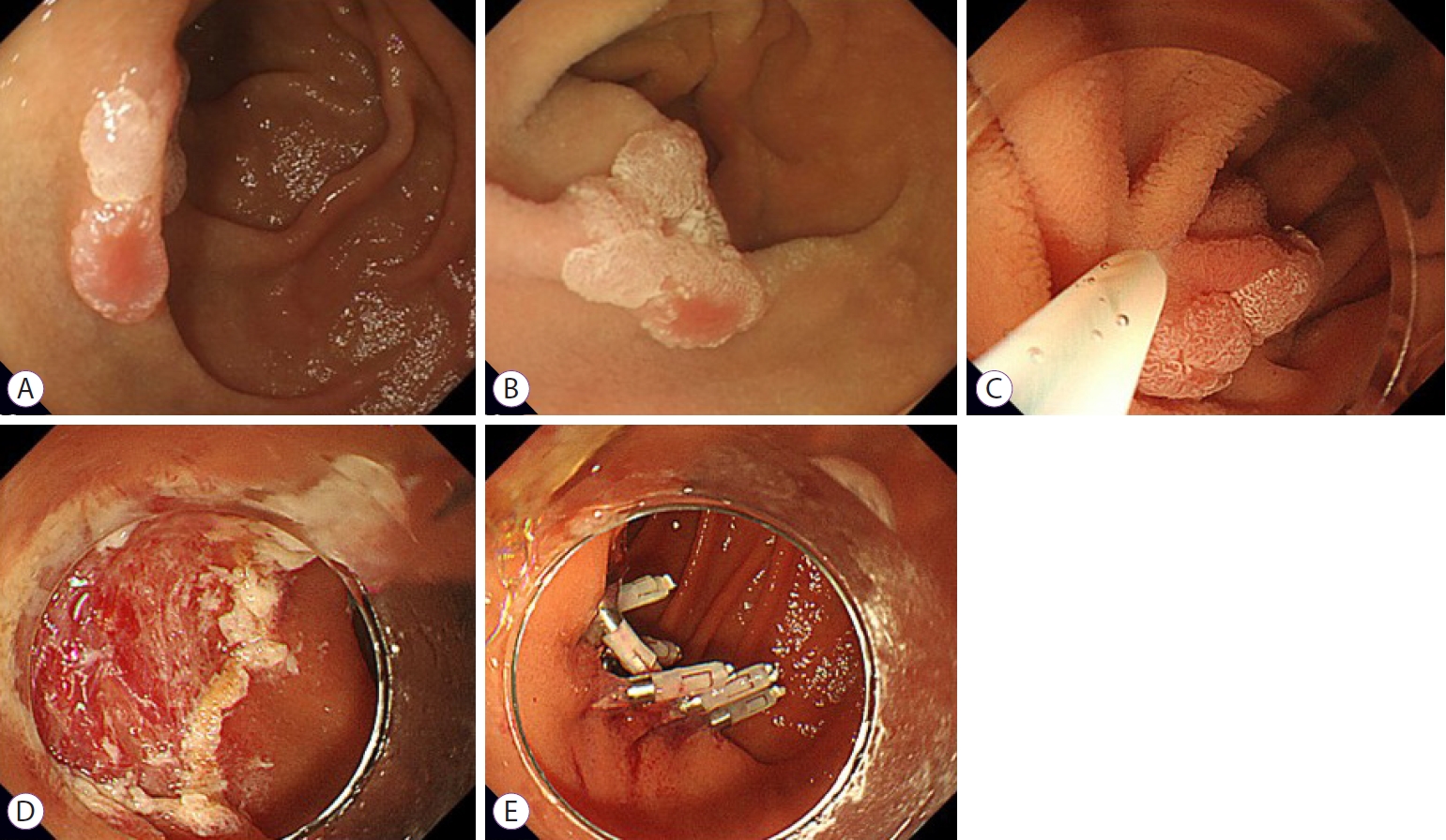
In contrast, after suctioning out air followed by injecting saline into the duodenal lumen, the duodenal lesion and the surrounding normal mucosa floated upward, lifting the surface of the lesion rather than a single point. Furthermore, tension in the duodenal mucosa is nearly nonexistent in the submerged condition. Therefore, the submerged mucosa was easier to grasp than the mucosa filled with air. Thus, we could easily snare the duodenal lesion in the underwater condition, achieving an excellent rate of en bloc resection with UEMR (Fig. 1).
Oka reported that en bloc resection for colorectal neoplasia reduced the local recurrence rate when compared with piecemeal resection [23]. Although the observation period was short, we found no cases of residual tumor in the UEMR group. However, two cases of residual tumor were found in the piecemeal CEMR group. Generally, the large number of piecemeal specimens makes it difficult to accurately determine the curability of the lesion regardless of the resection method used.
CEMR cases required a certain amount of time before adequate submucosal lifting and ER had to be performed with insufficient submucosal lifting for some patients. Filling the duodenal lumen with saline is a simple method that can be easily applied by injecting saline through the channel of the endoscope. This process creates a stable field similar to adequate submucosal lifting in the submerged condition, enabling endoscopic treatment. Hence, the resection time and the total procedure time for UEMR were shorter than those for CEMR in this study.
Binmoeller et al. reported that endoscopic ultrasonography in the underwater condition revealed a circular shape of the muscularis propria while maintaining native thickness [19]. Using the snaring procedure in this situation reduces the risk of grasping the muscle layer and facilitates conditions in which the lesion can be treated safely without intraoperative perforation. In addition, the underwater resection itself may reduce thermal damage to the duodenal wall.
It has been reported that complete closure of the post-resection wound prevents exposure to bile and pancreatic fluid and may also reduce postsurgical bleeding and delayed perforation [13,24,25]. Our study showed no significant difference in the success rate of prophylactic clipping between the UEMR group and the CEMR group. However, we could easily perform endoclip closure of the mucosal defect in all UEMR cases, since the surrounding mucosa after UEMR remained soft due to the lack of submucosal injection.
Kiguchi et al. reported the retrospective therapeutic results of UEMR and CEMR for SNADETs sized ≤20 mm [26]. The primary endpoint of the study was the resection rate without conversion to ESD. The conversion rate to ESD was significantly lower in UEMR than in CEMR. However, among the patients who underwent ER without conversion to ESD, UEMR cases had a significantly lower proportion of en bloc resection and R0 resection than CEMR cases [26]. In contrast, the primary endpoints of our study were en bloc resection rate and R0 resection rate of UEMR or CEMR. UEMR cases had a significantly higher proportion of en bloc resection and R0 resection than CEMR cases. No cases were converted to ESD in the present study. Particularly, UEMR showed remarkable usefulness for SNADETs of the “second part” and SNADETs sized ≤10 mm regardless of the lesion morphology. Duodenal ESD is one of the most challenging endoscopic procedures to perform in the digestive tract. UEMR is considered highly advantageous as an ER procedure for SNADET, since immediate change in the treatment strategy of difficult EMR cases to ESD requires a very advanced endoscopic technique.
While performing UEMR for SNADETs of the “second part” of the duodenum, the procedure might be difficult or rather time-consuming, as the water continues to go down without filling. In our experience, by evacuating the air in the stomach, the maneuverability of the endoscope is improved and it becomes easy to fill the second portion of the duodenum with water. Moreover, postural change is also useful in achieving good conditions for UEMR.
Our study has several limitations. It was a single-center retrospective study with a small number of patients. Moreover, the follow-up period was short. Hence, collection of long-term follow-up data is warranted.
In conclusion, UEMR is a safe and effective treatment for SNADET when compared with CEMR. Further large-scale multicenter studies are needed to confirm the effectiveness of UEMR.
We thank Dr. Yoji Takeuchi (Department of Gastrointestinal Oncology, Osaka International Cancer Institute, Osaka, Japan) for his insightful comments.
Fig. 1.
Underwater endoscopic mucosal resection for a duodenal intramucosal adenocarcinoma sized 11 mm. (A) Upper gastrointestinal endoscopy revealed an elevated lesion in the inferior duodenal angle. (B, C) The lesion floated up and became easy to snare in the submerged condition. (D) Ulcer after underwater endoscopic mucosal resection. (E) Complete closure with endoclips (EZ clip; Olympus, Tokyo, Japan).
ce-2020-147f1.jpg
Table 1.
Baseline Characteristics of Participants
UEMR (n=28) EMR (n=18) p-value
Age, yr 0.381
 Mean±SD 69.3±11.9 66.3±10.2
Sex 0.739
 Male, n (%) 20 (71.4) 14 (77.8)
Location, n (%) 0.562
 First part, n (%) 6 (21.4) 4 (22.2)
 Second part, n (%) 21 (75.0) 14 (77.8)
 Third part, n (%) 1 (3.6) 0 (0.0)
Location from the major papilla, n (%) 1.000
 Proximal to the major papilla, n (%) 10 (35.7) 6 (33.3)
 Distal to the major papilla, n (%) 18 (64.3) 12 (66.7)
Median (range) lesion size, (mm) 8.0 (2.0–20.0) 11.0 (3.0–20.0) 0.603
Macroscopic type, n (%) 0.439
 0-I 6 (21.4) 2 (11.1)
 0-IIa 15 (53.6) 13 (72.2)
 0-IIc 7 (25.0) 3 (16.7)

EMR, endoscopic mucosal resection; SD, standard deviation; UEMR, underwater endoscopic mucosal resection.

Table 2.
Treatment Outcomes
UEMR (n=28) EMR (n=18) p-value
Median (range) resection time, min 4.0 (2.0–11.0) 9.5 (4.0–57.0) 0.004a)
Median (range) total procedure time, min 13.0 (5.0–32.0) 19.0 (6.0–57.0) 0.013a)
En bloc resection, n (%) 27 (96.4) 13 (72.2) 0.028a)
R0 resection, n (%) 20 (71.4) 9 (50.0) 0.212
Prophylactic clipping, n (%) 28 (100.0) 17 (94.4) 0.391
Horizonal margin, n (%) 0.212
 Negative 20 (71.4) 9 (50.0)
 Positive or inconclusive 8 (28.6) 9 (50.0)
Vertical margin, n (%) 0.148
 Negative 28 (100.0) 16 (89.9)
 Positive or inconclusive 0 (0.0) 2 (11.1)
Pathological diagnosis, n (%) 0.346
 LGIN/Adenoma (Category 3) 13 (46.4) 6 (33.3)
 HGIN/Intramucosal adenocarcinoma (Category 4) 15 (53.6) 11 (61.1)
 Submucosal invasive cancer (Category 5) 0 (0.0) 1 (5.6)

EMR, endoscopic mucosal resection; HGIN, high grade intraepithelial neoplasia; LGIN, low grade intraepithelial neoplasia; UEMR, underwater endoscopic mucosal resection.

a)Statistically significant.

Table 3.
Detailed Outcomes of Each Endoscopic Resection Method
Location
First part UEMR (n=6) EMR (n=4) p-value
En bloc resection, n (%) 5 (83.3) 3 (75.0) 1.000
 R0 resection, n (%) 4 (66.7) 2 (50.0) 1.000
 Median (range) resection time, min 5.5 (3.0–19.0) 12.0 (8.0–57.0) 0.038a)
 Median (range) total procedure time, min 15.0 (8.0–28.0) 18.0 (13.0–57.0) 0.067
Second part UEMR (n=21) EMR (n=14) p-value
En bloc resection, n (%) 21 (100.0) 10 (71.4) 0.019a)
 R0 resection, n (%) 16 (76.2) 7 (50.0) 0.153
 Median (range) resection time, min 4.0 (3.0–10.0) 10.0 (4.0–15.0) 0.001a)
 Median (range) total procedure time, min 13.0 (5.0–32.0) 19.0 (4.0–35.0) 0.043a)

Size

≤10 mm UEMR (n=18) EMR (n=9) p-value
En bloc resection, n (%) 18 (100.0) 7 (77.8) 0.103
 R0 resection, n (%) 16 (88.9) 5 (55.6) 0.136
 Median (range) resection time, min 4.0 (2.0–11.0) 10.0 (4.0–15.0) 0.008a)
 Median (range) total procedure time, min 11.0 (5.0–15.0) 19.0 (4.0–35.0) 0.027a)
>10 mm UEMR (n=10) EMR (n=9) p-value
En bloc resection, n (%) 9 (90.0) 6 (66.7) 0.303
 R0 resection, n (%) 5 (50.0) 4 (44.4) 1.000
 Median (range) resection time, min 6.5 (3.0–19.0) 12.0 (4.0–57.0) 0.210
 Median (range) total procedure time, min 19.5 (10.0–32.0) 23.0 (7.0–57.0) 0.556

Morophology

Elevated type UEMR (n=21) EMR (n=15) p-value
En bloc resection, n (%) 21 (100.0) 11 (73.3) 0.138
 R0 resection, n (%) 17 (81.0) 8 (53.3) 0.141
 Median (range) resection time, min 6.0 (3.0–19.0) 9.0 (4.0–57.0) 0.012a)
 Median (range) total procedure time, min 13.0 (5.0–32.0) 17.0 (4.0–57.0) 0.125
Depressed type UEMR (n=7) EMR (n=3) p-value
En bloc resection, n (%) 7 (100.0) 2 (66.7) 0.300
 R0 resection, n (%) 4 (57.1) 1 (33.3) 1.000
 Median (range) resection time, min 4.0 (2.0–5.0) 12.0 (12.0–15.0) 0.014a)
 Median (range) total procedure time, min 14.0 (5.0–18.0) 22.0 (21.0–30.0) 0.016a)

EMR, endoscopic mucosal resection; UEMR, underwater endoscopic mucosal resection.

a)Statistically significant.

Table 4.
Adverse Events and Follow-up Outcome
UEMR (n=28) EMR (n=18) p-value
Delayed bleeding, n (%) 0 (0.0) 0 (0.0) 1.000
Perforation, n (%) 0 (0.0) 0 (0.0) 1.000
Recurrence, n (%) 0 (0.0) 2 (11.1) 0.148

EMR, endoscopic mucosal resection; UEMR, underwater endoscopic mucosal resection.

  • 1. Höchter W, Weingart J, Seib HJ, Ottenjann R. [Duodenal polyps. Incidence, histologic substrate and significance]. Dtsch Med Wochenschr 1984;109:1183–1186.ArticlePubMed
  • 2. Jepsen JM, Persson M, Jakobsen NO, et al. Prospective study of prevalence and endoscopic and histopathologic characteristics of duodenal polyps in patients submitted to upper endoscopy. Scand J Gastroenterol 1994;29:483–487.ArticlePubMed
  • 3. Murray MA, Zimmerman MJ, Ee HC. Sporadic duodenal adenoma is associated with colorectal neoplasia. Gut 2004;53:261–265.ArticlePubMedPMC
  • 4. Jung SH, Chung WC, Kim EJ, et al. Evaluation of non-ampullary duodenal polyps: comparison of non-neoplastic and neoplastic lesions. World J Gastroenterol 2010;16:5474–5480.ArticlePubMedPMC
  • 5. Goda K, Kikuchi D, Yamamoto Y, et al. Endoscopic diagnosis of superficial non-ampullary duodenal epithelial tumors in Japan: multicenter case series. Dig Endosc 2014;26(Suppl 2):23–29.Article
  • 6. Seifert E, Schulte F, Stolte M. Adenoma and carcinoma of the duodenum and papilla of Vater: a clinicopathologic study. Am J Gastroenterol 1992;87:37–42.PubMed
  • 7. Okada K, Fujisaki J, Kasuga A, et al. Sporadic nonampullary duodenal adenoma in the natural history of duodenal cancer: a study of follow-up surveillance. Am J Gastroenterol 2011;106:357–364.ArticlePubMed
  • 8. Asbun HJ. Management of duodenal polyps in the era of maximal interventional endoscopy and minimally invasive surgery: a surgical perspective. Gastrointest Endosc 2016;84:697–699.ArticlePubMed
  • 9. Friedrich-Rust M, Ell C. Early-stage small-bowel adenocarcinoma: a review of local endoscopic therapy. Endoscopy 2005;37:755–759.ArticlePubMed
  • 10. Nonaka S, Oda I, Tada K, et al. Clinical outcome of endoscopic resection for nonampullary duodenal tumors. Endoscopy 2015;47:129–135.ArticlePubMed
  • 11. Apel D, Jakobs R, Spiethoff A, Riemann JF. Follow-up after endoscopic snare resection of duodenal adenomas. Endoscopy 2005;37:444–448.ArticlePubMed
  • 12. Alexander S, Bourke MJ, Williams SJ, Bailey A, Co J. EMR of large, sessile, sporadic nonampullary duodenal adenomas: technical aspects and long-term outcome (with videos). Gastrointest Endosc 2009;69:66–73.ArticlePubMed
  • 13. Maruoka D, Arai M, Kishimoto T, et al. Clinical outcomes of endoscopic resection for nonampullary duodenal high-grade dysplasia and intramucosal carcinoma. Endoscopy 2013;45:138–141.ArticlePubMed
  • 14. Klein A, Nayyar D, Bahin FF, et al. Endoscopic mucosal resection of large and giant lateral spreading lesions of the duodenum: success, adverse events, and long-term outcomes. Gastrointest Endosc 2016;84:688–696.ArticlePubMed
  • 15. Jung JH, Choi KD, Ahn JY, et al. Endoscopic submucosal dissection for sessile, nonampullary duodenal adenomas. Endoscopy 2013;45:133–135.ArticlePubMed
  • 16. Matsumoto S, Miyatani H, Yoshida Y. Endoscopic submucosal dissection for duodenal tumors: a single-center experience. Endoscopy 2013;45:136–137.ArticlePubMed
  • 17. Hoteya S, Yahagi N, Iizuka T, et al. Endoscopic submucosal dissection for nonampullary large superficial adenocarcinoma/adenoma of the duodenum: feasibility and long-term outcomes. Endosc Int Open 2013;1:2–7.ArticlePubMedPMC
  • 18. Inoue T, Uedo N, Yamashina T, et al. Delayed perforation: a hazardous complication of endoscopic resection for non-ampullary duodenal neoplasm. Dig Endosc 2014;26:220–227.ArticlePubMed
  • 19. Binmoeller KF, Shah JN, Bhat YM, Kane SD. “Underwater” EMR of sporadic laterally spreading nonampullary duodenal adenomas (with video). Gastrointest Endosc 2013;78:496–502.ArticlePubMed
  • 20. Yamasaki Y, Uedo N, Takeuchi Y, et al. Underwater endoscopic mucosal resection for superficial nonampullary duodenal adenomas. Endoscopy 2018;50:154–158.ArticlePubMed
  • 21. Dixon MF. Gastrointestinal epithelial neoplasia: Vienna revisited. Gut 2002;51:130–131.ArticlePubMedPMC
  • 22. Kinoshita S, Nishizawa T, Ochiai Y, et al. Accuracy of biopsy for the preoperative diagnosis of superficial nonampullary duodenal adenocarcinoma. Gastrointest Endosc 2017;86:329–332.ArticlePubMed
  • 23. Oka S, Tanaka S, Saito Y, et al. Local recurrence after endoscopic resection for large colorectal neoplasia: a multicenter prospective study in Japan. Am J Gastroenterol 2015;110:697–707.ArticlePubMed
  • 24. Hoteya S, Kaise M, Iizuka T, et al. Delayed bleeding after endoscopic submucosal dissection for non-ampullary superficial duodenal neoplasias might be prevented by prophylactic endoscopic closure: analysis of risk factors. Dig Endosc 2015;27:323–330.ArticlePubMed
  • 25. Yahagi N, Kato M, Ochiai Y, et al. Outcomes of endoscopic resection for superficial duodenal epithelial neoplasia. Gastrointest Endosc 2018;88:676–682.ArticlePubMed
  • 26. Kiguchi Y, Kato M, Nakayama A, et al. Feasibility study comparing underwater endoscopic mucosal resection and conventional endoscopic mucosal resection for superficial non-ampullary duodenal epithelial tumor < 20 mm. Dig Endosc 2020;32:753–760.ArticlePubMed

Figure & Data

REFERENCES

    Citations

    Citations to this article as recorded by  
    • Underwater Endoscopic Mucosal Resection Vs Conventional Endoscopic Mucosal Resection for Superficial Nonampullary Duodenal Epithelial Tumors in the Western Setting
      Rui Morais, José Amorim, Renato Medas, Bernardo Sousa-Pinto, João Santos-Antunes, Romain Legros, Jérémie Albouys, Frédéric Moll, Margarida Marques, Filipe Vilas-Boas, Eduardo Rodrigues-Pinto, Irene Gullo, Fátima Carneiro, Elisa Gravito Soares, Pedro Amaro
      Clinical Gastroenterology and Hepatology.2025; 23(1): 79.     CrossRef
    • Conventional versus underwater endoscopic resection for superficial non-ampullary duodenal epithelial tumours
      Hajime Miyazaki, Osamu Dohi, Tsugitaka Ishida, Mayuko Seya, Katsuma Yamauchi, Hayato Fukui, Takeshi Yasuda, Takuma Yoshida, Naoto Iwai, Toshifumi Doi, Ryohei Hirose, Ken Inoue, Akihito Harusato, Naohisa Yoshida, Kazuhiko Uchiyama, Tomohisa Takagi, Takeshi
      Japanese Journal of Clinical Oncology.2024; 54(2): 137.     CrossRef
    • Can underwater endoscopic mucosal resection be an alternative to conventional endoscopic mucosal resection for superficial non‐ampullary duodenal epithelial tumors?
      Hidenori Tanaka, Yuji Urabe, Hiroki Takemoto, Kazuki Ishibashi, Hirona Konishi, Yuka Matsubara, Yudai Takehara, Shin Morimoto, Fumiaki Tanino, Noriko Yamamoto, Hajime Teshima, Junichi Mizuno, Issei Hirata, Hirosato Tamari, Akiyoshi Tsuboi, Ken Yamashita,
      DEN Open.2024;[Epub]     CrossRef
    • Underwater Techniques in Gastrointestinal Endoscopy: Diving into the Depths
      Sandro Sferrazza, Giulio Calabrese, Roberta Maselli, Rui Morais, Antonio Facciorusso, Georgios Mavrogenis, Roberto Di Mitri, Alessandro Repici, Marcello Maida
      Cancers.2024; 16(20): 3535.     CrossRef
    • Underwater versus conventional endoscopic mucosal resection for superficial non-ampullary duodenal epithelial tumors ≤20mm: A systematic review and meta-analysis
      Xiu-He Lv, Rong Luo, Qing Lu, Kai Deng, Jin-Lin Yang
      Digestive and Liver Disease.2023; 55(6): 714.     CrossRef
    • Underwater Versus Conventional Endoscopic Mucosal Resection for Superficial Non-ampullary Duodenal Epithelial Tumors: A Systematic Review and Meta-Analysis
      Jae Gon Lee, Sang Pyo Lee, Hyun Joo Jang, Sea Hyub Kae
      Digestive Diseases and Sciences.2023; 68(4): 1482.     CrossRef
    • A feasibility study comparing gel immersion endoscopic resection and underwater endoscopic mucosal resection for superficial nonampullary duodenal epithelial tumors
      Akihiro Miyakawa, Toshio Kuwai, Yukie Sakuma, Manabu Kubota, Akira Nakamura, Ei Itobayashi, Haruhisa Shimura, Yoshio Suzuki, Kenji Shimura
      Endoscopy.2023; 55(03): 261.     CrossRef
    • Underwater Endoscopic Mucosal Resection Versus Conventional Endoscopic Mucosal Resection for Superficial Non-ampullary Duodenal Epithelial Tumors ≤20 mm
      Zhikun Yin, Ji Li, Weilin Yang, Weifeng Huang, Dong Xu, Xiaoyi Lei, Jinyan Zhang
      Journal of Clinical Gastroenterology.2023; 57(9): 928.     CrossRef
    • Efficacy and safety of underwater endoscopic mucosal resection for ≤20 mm superficial non-ampullary duodenal epithelial tumors: Systematic review and meta-analysis
      Jixiang Liu, Shaojie Duan, Yichong Wang, Hongye Peng, Youjia Kong, Shukun Yao
      Frontiers in Medicine.2023;[Epub]     CrossRef
    • Underwater versus conventional EMR for nonpedunculated colorectal lesions: a randomized clinical trial
      Luciano Lenz, Bruno Martins, Gustavo Andrade de Paulo, Fabio Shiguehissa Kawaguti, Elisa Ryoka Baba, Ricardo Sato Uemura, Carla Cristina Gusmon, Sebastian Naschold Geiger, Renata Nobre Moura, Caterina Pennacchi, Marcelo Simas de Lima, Adriana Vaz Safatle-
      Gastrointestinal Endoscopy.2023; 97(3): 549.     CrossRef
    • Endoscopic resection of superficial non‐ampullary duodenal epithelial tumor
      Motohiko Kato, Takanori Kanai, Naohisa Yahagi
      DEN Open.2022;[Epub]     CrossRef
    • The Application of Underwater Endoscopic Mucosal Resection for Nonampullary Duodenal Adenomas
      Xiu-He Lv, Jin-Lin Yang
      Clinical Gastroenterology and Hepatology.2022; 20(8): 1884.     CrossRef
    • Utility of underwater EMR for nonpolypoid superficial nonampullary duodenal epithelial tumors ≤20 mm
      Kenichiro Okimoto, Daisuke Maruoka, Tomoaki Matsumura, Kengo Kanayama, Naoki Akizue, Yuki Ohta, Takashi Taida, Keiko Saito, Yosuke Inaba, Yohei Kawasaki, Makoto Arai, Jun Kato, Naoya Kato
      Gastrointestinal Endoscopy.2022; 95(1): 140.     CrossRef
    • Reply
      Yasushi Yamasaki, Noriya Uedo
      Clinical Gastroenterology and Hepatology.2022; 20(8): 1884.     CrossRef
    • Appropriate selection of endoscopic resection for superficial nonampullary duodenal adenomas in association with recurrence
      Kenichiro Okimoto, Daisuke Maruoka, Tomoaki Matsumura, Kengo Kanayama, Naoki Akizue, Yuki Ohta, Takashi Taida, Keiko Saito, Yosuke Inaba, Yohei Kawasaki, Jun Kato, Naoya Kato
      Gastrointestinal Endoscopy.2022; 95(5): 939.     CrossRef
    • Reply to Lv and Yang
      Motohiko Kato, Yoji Takeuchi, Shu Hoteya, Tsuneo Oyama, Satoru Nonaka, Shoichi Yoshimizu, Naomi Kakushima, Ken Ohata, Hironori Yamamoto, Yuko Hara, Hisashi Doyama, Osamu Dohi, Yasushi Yamasaki, Hiroya Ueyama, Kengo Takimoto, Koichi Kurahara, Tomoaki Tashi
      Endoscopy.2022; 54(05): 523.     CrossRef
    • Endoscopic treatment selection for superficial duodenal tumors: pay attention to small lesions
      Xiu-He Lv, Jin-Lin Yang
      Endoscopy.2022; 54(05): 522.     CrossRef
    • Resectability of underwater endoscopic mucosal resection for duodenal tumor: A single‐center, retrospective pilot study
      Yosuke Toya, Masaki Endo, Masanao Yamazato, Shun Yamada, Tomo Kumei, Minami Hirai, Makoto Eizuka, Toshifumi Morishita, Risaburo Akasaka, Shunichi Yanai, Noriyuki Uesugi, Tamotsu Sugai, Takayuki Matsumoto
      Journal of Gastroenterology and Hepatology.2021; 36(11): 3191.     CrossRef

    • PubReader PubReader
    • ePub LinkePub Link
    • Cite
      CITE
      export Copy Download
      Close
      Download Citation
      Download a citation file in RIS format that can be imported by all major citation management software, including EndNote, ProCite, RefWorks, and Reference Manager.

      Format:
      • RIS — For EndNote, ProCite, RefWorks, and most other reference management software
      • BibTeX — For JabRef, BibDesk, and other BibTeX-specific software
      Include:
      • Citation for the content below
      Efficacy of Underwater Endoscopic Mucosal Resection for Superficial Non-Ampullary Duodenal Epithelial Tumor
      Clin Endosc. 2021;54(3):371-378.   Published online February 18, 2021
      Close
    • XML DownloadXML Download
    Figure
    • 0
    Related articles
    Efficacy of Underwater Endoscopic Mucosal Resection for Superficial Non-Ampullary Duodenal Epithelial Tumor
    Image
    Fig. 1. Underwater endoscopic mucosal resection for a duodenal intramucosal adenocarcinoma sized 11 mm. (A) Upper gastrointestinal endoscopy revealed an elevated lesion in the inferior duodenal angle. (B, C) The lesion floated up and became easy to snare in the submerged condition. (D) Ulcer after underwater endoscopic mucosal resection. (E) Complete closure with endoclips (EZ clip; Olympus, Tokyo, Japan).
    Efficacy of Underwater Endoscopic Mucosal Resection for Superficial Non-Ampullary Duodenal Epithelial Tumor
    UEMR (n=28) EMR (n=18) p-value
    Age, yr 0.381
     Mean±SD 69.3±11.9 66.3±10.2
    Sex 0.739
     Male, n (%) 20 (71.4) 14 (77.8)
    Location, n (%) 0.562
     First part, n (%) 6 (21.4) 4 (22.2)
     Second part, n (%) 21 (75.0) 14 (77.8)
     Third part, n (%) 1 (3.6) 0 (0.0)
    Location from the major papilla, n (%) 1.000
     Proximal to the major papilla, n (%) 10 (35.7) 6 (33.3)
     Distal to the major papilla, n (%) 18 (64.3) 12 (66.7)
    Median (range) lesion size, (mm) 8.0 (2.0–20.0) 11.0 (3.0–20.0) 0.603
    Macroscopic type, n (%) 0.439
     0-I 6 (21.4) 2 (11.1)
     0-IIa 15 (53.6) 13 (72.2)
     0-IIc 7 (25.0) 3 (16.7)
    UEMR (n=28) EMR (n=18) p-value
    Median (range) resection time, min 4.0 (2.0–11.0) 9.5 (4.0–57.0) 0.004a)
    Median (range) total procedure time, min 13.0 (5.0–32.0) 19.0 (6.0–57.0) 0.013a)
    En bloc resection, n (%) 27 (96.4) 13 (72.2) 0.028a)
    R0 resection, n (%) 20 (71.4) 9 (50.0) 0.212
    Prophylactic clipping, n (%) 28 (100.0) 17 (94.4) 0.391
    Horizonal margin, n (%) 0.212
     Negative 20 (71.4) 9 (50.0)
     Positive or inconclusive 8 (28.6) 9 (50.0)
    Vertical margin, n (%) 0.148
     Negative 28 (100.0) 16 (89.9)
     Positive or inconclusive 0 (0.0) 2 (11.1)
    Pathological diagnosis, n (%) 0.346
     LGIN/Adenoma (Category 3) 13 (46.4) 6 (33.3)
     HGIN/Intramucosal adenocarcinoma (Category 4) 15 (53.6) 11 (61.1)
     Submucosal invasive cancer (Category 5) 0 (0.0) 1 (5.6)
    Location
    First part UEMR (n=6) EMR (n=4) p-value
    En bloc resection, n (%) 5 (83.3) 3 (75.0) 1.000
     R0 resection, n (%) 4 (66.7) 2 (50.0) 1.000
     Median (range) resection time, min 5.5 (3.0–19.0) 12.0 (8.0–57.0) 0.038a)
     Median (range) total procedure time, min 15.0 (8.0–28.0) 18.0 (13.0–57.0) 0.067
    Second part UEMR (n=21) EMR (n=14) p-value
    En bloc resection, n (%) 21 (100.0) 10 (71.4) 0.019a)
     R0 resection, n (%) 16 (76.2) 7 (50.0) 0.153
     Median (range) resection time, min 4.0 (3.0–10.0) 10.0 (4.0–15.0) 0.001a)
     Median (range) total procedure time, min 13.0 (5.0–32.0) 19.0 (4.0–35.0) 0.043a)

    Size

    ≤10 mm UEMR (n=18) EMR (n=9) p-value
    En bloc resection, n (%) 18 (100.0) 7 (77.8) 0.103
     R0 resection, n (%) 16 (88.9) 5 (55.6) 0.136
     Median (range) resection time, min 4.0 (2.0–11.0) 10.0 (4.0–15.0) 0.008a)
     Median (range) total procedure time, min 11.0 (5.0–15.0) 19.0 (4.0–35.0) 0.027a)
    >10 mm UEMR (n=10) EMR (n=9) p-value
    En bloc resection, n (%) 9 (90.0) 6 (66.7) 0.303
     R0 resection, n (%) 5 (50.0) 4 (44.4) 1.000
     Median (range) resection time, min 6.5 (3.0–19.0) 12.0 (4.0–57.0) 0.210
     Median (range) total procedure time, min 19.5 (10.0–32.0) 23.0 (7.0–57.0) 0.556

    Morophology

    Elevated type UEMR (n=21) EMR (n=15) p-value
    En bloc resection, n (%) 21 (100.0) 11 (73.3) 0.138
     R0 resection, n (%) 17 (81.0) 8 (53.3) 0.141
     Median (range) resection time, min 6.0 (3.0–19.0) 9.0 (4.0–57.0) 0.012a)
     Median (range) total procedure time, min 13.0 (5.0–32.0) 17.0 (4.0–57.0) 0.125
    Depressed type UEMR (n=7) EMR (n=3) p-value
    En bloc resection, n (%) 7 (100.0) 2 (66.7) 0.300
     R0 resection, n (%) 4 (57.1) 1 (33.3) 1.000
     Median (range) resection time, min 4.0 (2.0–5.0) 12.0 (12.0–15.0) 0.014a)
     Median (range) total procedure time, min 14.0 (5.0–18.0) 22.0 (21.0–30.0) 0.016a)
    UEMR (n=28) EMR (n=18) p-value
    Delayed bleeding, n (%) 0 (0.0) 0 (0.0) 1.000
    Perforation, n (%) 0 (0.0) 0 (0.0) 1.000
    Recurrence, n (%) 0 (0.0) 2 (11.1) 0.148
    Table 1. Baseline Characteristics of Participants

    EMR, endoscopic mucosal resection; SD, standard deviation; UEMR, underwater endoscopic mucosal resection.

    Table 2. Treatment Outcomes

    EMR, endoscopic mucosal resection; HGIN, high grade intraepithelial neoplasia; LGIN, low grade intraepithelial neoplasia; UEMR, underwater endoscopic mucosal resection.

    Statistically significant.

    Table 3. Detailed Outcomes of Each Endoscopic Resection Method

    EMR, endoscopic mucosal resection; UEMR, underwater endoscopic mucosal resection.

    Statistically significant.

    Table 4. Adverse Events and Follow-up Outcome

    EMR, endoscopic mucosal resection; UEMR, underwater endoscopic mucosal resection.


    Clin Endosc : Clinical Endoscopy Twitter Facebook
    Close layer
    TOP