INTRODUCTION
Esophageal cancer ranks seventh in terms of incidence and is the sixth most common cause of cancer-related mortality worldwide [1]. Esophageal squamous cell carcinoma (ESCC) is the most common histological type of esophageal cancer [2-4], and the incidence of esophageal adenocarcinoma (EAC) is increasing, especially in Western countries [5]. Esophageal dysplasia, including low-grade intraepithelial neoplasia, high-grade intraepithelial neoplasia (HGIN), and BarrettŌĆÖs esophagus (BE) are the precursor lesions of ESCC and EAC [6,7].
Despite the improvement in treatment modalities, the 5-year survival of esophageal cancer is between 10% and 25% and the mortality rate is still high due to an advanced stage at diagnosis [4,8,9]. However, the prognosis can be improved up to 95% when it is detected and treated as an early-stage disease that can be treated with endoscopic resection [10]. Predicting the depth of the invasion of early esophageal cancer is important to determine a proper candidate lesion for endoscopic resection since the invasion depth has a reliable relationship with the rate of lymph node metastasis [11-13]. The 5-year cause-specific survival and 5-year overall survival rates were reported to be 98ŌĆō100% and 85ŌĆō95% in endoscopic submucosal dissection (ESD) of the depth of the epithelium (EP) or lamina propria mucosae (LPM), respectively [14].
White-light endoscopy (WLE) is the standard modality for detecting esophageal neoplastic lesions. However, the endoscopic features of early esophageal neoplastic lesions under WLE are subtle and isochromatic [15,16]. In addition, BE-related dysplasia or early EAC is prone to remain undetected when using standard WLE since these lesions are flat and unremarkable [17]. A multicenter randomized controlled trial (RCT) found that the sensitivity of WLE for superficial ESCC was less than 60% [15]. Image-enhanced endoscopy (IEE), including dye-based chromoendoscopy and virtual chromoendoscopy with or without magnifying endoscopy (ME) is a new diagnostic endoscopic technique that has been helpful in the detection of tumors and predicting the depth of invasion of esophageal cancer. This review aimed to provide an overview of the current status and advancements of IEE-related technologies for the early detection of esophageal neoplasms.
CONVENTIONAL CHROMOENDOSCOPY
Conventional chromoendoscopy is a technique that enhances the mucosal structural details by spraying dyes onto mucosal surfaces. It assists in distinguishing the abnormal mucosa from the normal mucosa by contrast to the mucosal surface by permeating depressed mucosal folds or grooves (i.e., contrast dye) or interaction with specific intracellular materials or elements (i.e., absorptive dye).
LugolŌĆÖs iodine
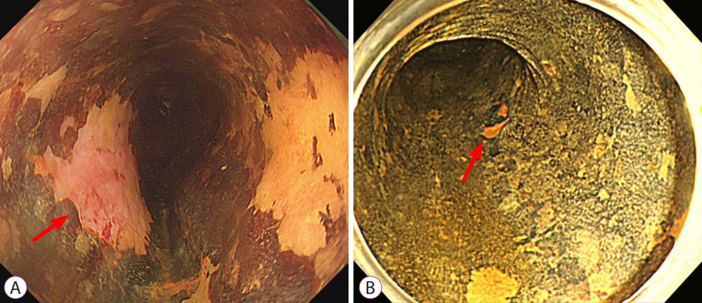
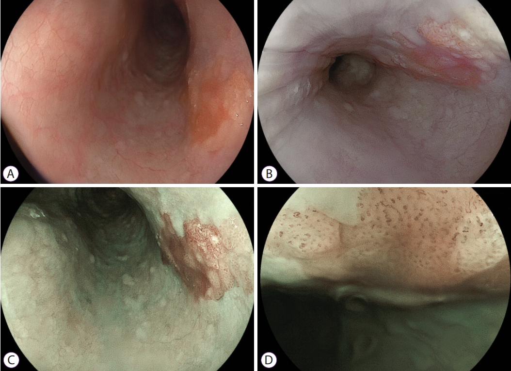
LugolŌĆÖs iodine is the most widely used absorptive dye for detecting esophageal squamous cell dysplasia and ESCC. Mature esophageal squamous epithelium contains abundant glycogen, which is stained a dark brown color with iodine; however, the dysplastic lesion exhibits either decreased or absent glycogen content as well as demonstrates a characteristic of unstained appearance on inspection following application of LugolŌĆÖs iodine. The unstained area of LugolŌĆÖs iodine is useful to detect esophageal squamous cell dysplasia or ESCC [18]. LugolŌĆÖs chromoendoscopy (LCE) is the gold standard for the detection of ESCC. LCE can help identify the borders of esophageal cancer and facilitate precise biopsies. The European Society of Gastroenterology Endoscopy guidelines recommend that it should be used to demarcate the lateral margins of superficial tumors and to identify synchronous esophageal lesions [19]. One prospective observational study investigated the prevalence of esophageal cancer in patients with primary head and neck cancer using LCE and WLE. It has been shown that the diagnosis of advanced and invasive esophageal cancer was equivalent in both modalities; however, the LCE can detect HGIN more precisely than WLE (100% vs. 55 %) [20]. The sensitivity of LCE for the diagnosis of squamous cell dysplasia and ESCC ranges from 91% to 100% [18,21-24]. It would hard to be stained by LugolŌĆÖs Iodine after changing into HGIN and ESCC due to lack of cells containing glycogen. Therefore, these lesions are observed with a pinkish color after the brown color of the iodine solution has faded [25]. The pink-color discoloration within the Lugol-voiding area observed after 2 to 3 minutes from the staining (i.e. pink-color sign, Fig. 1) has been significantly correlated with HGIN and ESCC on histology (sensitivity was 91.9% and specificity was 94.0%) [25]. Nevertheless, LCE increases the duration of endoscopic examination and can cause mucosal irritation leading to adverse events, such as allergic reaction, retrosternal pain, and esophageal erosion or ulcers [26,27]. Occasionally, subtle early neoplastic lesions could temporarily disappear because of mucosal inflammation and interfere with endoscopic treatment for days to weeks [28]. A recent double-blind RCT, which aimed to evaluate patient discomfort with different iodine concentrations found that a 1% iodine solution leads to significantly lesser pain than a 2% iodine solution (p=0.02) with non-inferior visibility [29].
Acetic acid
Acetic acid (AA) is a useful dye for the early identification of dysplastic BE lesions. AA improves the visualization of the mucosal surface via reversible acetylation of nuclear proteins. The reaction only lasts for a few minutes, and dysplastic tissues lose the acetowhitening quicker than surrounding intestinal-type columnar metaplasia [30,31]. Two large trials have reported the effectiveness of AA in detecting neoplasms in BE in a high-risk population [31,32]. Additionally, one retrospective large cohort study showed that AA-guided biopsies significantly improved the detection rate of neoplasia (2% vs. 12.5%, p=0.0001) and requires 15 times fewer biopsies on per-biopsy analysis (0.025 vs. 0.0017, p=0.000) [33]. Recent meta-analyses showed that targeted biopsies with AA are useful for detecting HGIN and early adenocarcinoma of BE (sensitivity of 92% and specificity of 96%, respectively) [34,35].
Methylene blue and indigo carmine
Methylene blue selectively stains the specialized intestinal epithelium. Three randomized cross-over trials have shown that the diagnostic accuracy of methylene blue-assisted biopsies is higher than that of stepwise four-quadrant biopsies (ŌĆśSeattle protocolŌĆÖ) [36-38]. However, a meta-analysis of nine studies determined that methylene blue target biopsies were not superior to random biopsies for the diagnosis of dysplasia [39].
One of the contrast dyes, indigo carmine, seeps between the grooves and assists in observing the contour of the mucosal surface. It has been shown to be useful for the detection of colon adenomas. However, a randomized crossover study showed that indigo carmine did not increase the BEŌĆÖs dysplasia detection rate compared to high-resolution WLE [40].
VIRTUAL CHROMOENDOSCOPY
Virtual chromoendoscopy is a hardware-based technique. In contrast to conventional chromoendoscopy, it is easy to obtain an enhanced image by turning on a button-switch and allowing the colorimetric manipulation of target lesions and is replacing dye-based chromoendoscopy. Virtual chromoendoscopy can be divided into several categories. Light filter technologies using optical filters to adapt to specific wave-lengths include narrow-band imaging (NBI), which is the most investigated technique in the IEE field. Software-based digital image processing techniques include flexible spectral imaging color enhancement (FICE) and i-SCAN. The most recently introduced techniques, namely, blue laser imaging (BLI) and light color imaging (LCI) have similar principles in generating an endoscopic image to NBI. However, BLI and LCI use monochromatic lasers instead of xenon light with an optical filter.
NBI, with or without magnification
a. Esophageal squamous cell dysplasia and carcinoma
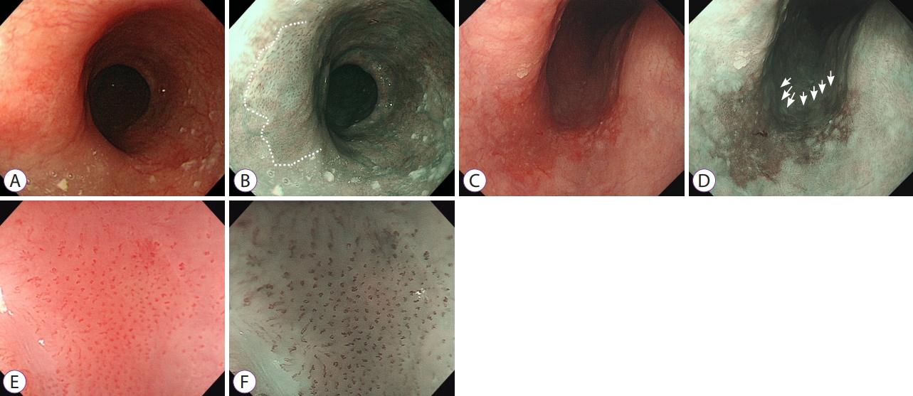
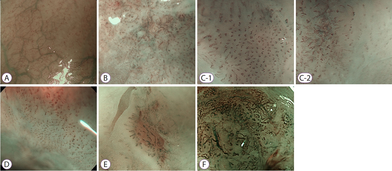
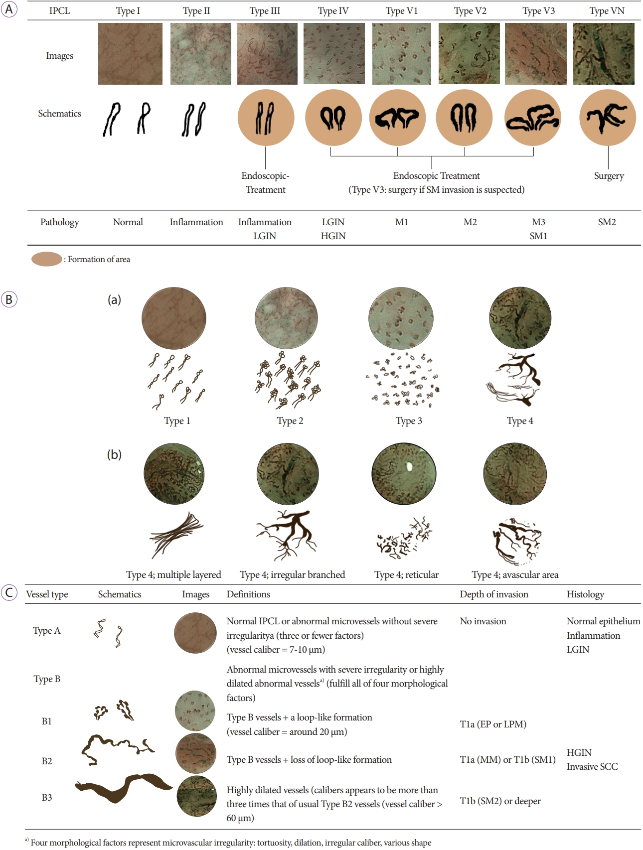
NBI is the most widely studied and used virtual chromoendoscopy. It utilizes two kinds of light via an optical filter: blue light (wavelength of 415┬▒30 nm) and green light (wavelength 540┬▒30 nm). The NBI system emphasizes capillary vessels by absorbing hemoglobin in capillaries located on the mucosal surface. Esophageal dysplasia and ESCC are brown in color under NBI (Fig. 2). The diagnostic accuracy of non-magnifying endoscopy with NBI (NM-NBI) for superficial ESCC was significantly higher than that of WLE [15]. A recent meta-analysis including 12 studies with 1,911 patients reported that NBI was suitable for esophageal evaluation to diagnose esophageal squamous cell neoplasm. NBI has been shown to be superior to LCE in the differentiation of neoplastic lesions from other benign mucosal alterations (sensitivity 88% vs. 92%, p>0.05; specificity 88% vs. 82%, p<0.001) [41]. A prospective comparative study of NM-NBI and LCE in screening early ESCC and HGIN found the accuracy and specificity of NM-NBI were superior to those of LCE (77.0% vs. 68.0%, p=0.03; 95.2% vs. 64.0%, p=0.01) [42]. NM-NBI could improve the early detection of ESCC; however, it could also yield false-positive results because the non-specific inflammatory lesions may be identified as ŌĆ£brownish areaŌĆØ [43]. ME can provide magnification of up to ├Ś150. This aids the diagnosis through a detailed examination of microvascular and microsurface features and helps to predict the depth of invasion of early esophageal neoplastic lesions when used with NBI [44]. ME-NBI is presumed to be useful and might reduce the false-positive rate of NM-NBI in the diagnosis of superficial ESCC [43]. In a multicenter, prospective RCT, MN-NBI detected superficial cancers more frequently than WLE in the esophagus (97% vs. 55%, p<0.001) [15]. A non-inferiority RCT that compared the diagnostic accuracy of ME-NBI with that of LCE showed no significant difference between the two modalities with respect to the sensitivity and in any other diagnostic accuracy measure for superficial ESCC detection [16]. Intrapapillary capillary loop (IPCL) patterns, which are well observed with ME-NBI, are useful for the early and accurate diagnosis of esophageal cell neoplasm and prediction of the depth of invasion of ESCC (Fig. 3). The IPCL is an intrapapillary capillary that arises from the fourth branch of the arborescent vessels into the epithelial papillae and forms single loops. IPCL patterns undergo stepwise morphologic changes of ŌĆ£dilationŌĆØ, ŌĆ£tortuosityŌĆØ, ŌĆ£change in caliberŌĆØ and ŌĆ£various shapesŌĆØ with the progression of esophageal cell neoplasm (Fig. 4) [45]. Inoue classification and Arima classification were proposed to classify the invasion depth of superficial ESCC based on the change in the microvascular pattern (Figs. 5A, 5B) [45-48]. The overall accuracy of IPCL-IV and IPCL-V was 80% after analyzing the histological correlation of the IPCL classification in 185 superficial ESCC [46]. Additionally, the sensitivity and specificity of ME-NBI to differentiate intramucosal cancer from submucosal cancer was 78% and 95%, respectively [49]. Although Inoue and Arima classifications have been used widely in routine clinical practice, the existence of two classifications has caused considerable confusion [50]. Recently, the Japan Esophageal Society proposed more simplified classification guidelines for ME diagnosis of ESCC (JES classification) based on the vascular architecture (Fig. 5C). The overall accuracy of the JES classification for the prediction of tumor invasion depth of type B micro-vessels was 90.5% [51]. One retrospective study showed reliable interobserver agreement of the JES classification (k=0.86, 95% confidence interval [CI]: 0.76ŌĆō0.95) [52]. A retrospective study of 256 superficial ESCC patients who underwent non-magnifying WLE and ME-NBI for the assessment of the invasion depth before ESD showed that the JES classification provided an accurate estimation of the invasion depth in patients who had superficial ESCC with a histopathological diagnosis of the EP/LPM invasion (positive predictive value of 93%) [50].
In fact, most of these studies were conducted in a tertiary hospital setting with expert endoscopists. Moreover, ME-NBI is still not very popular even in Japan, and it is not currently available in several non-expert settings. However, since early esophageal neoplastic lesions have no prominent symptoms, there is a greater chance of detection during screening or surveillance endoscopy in general practice with non-expert settings. One prospective, non-randomized study compared the diagnostic yield of NBI endoscopy for the screening of squamous HGIN between experienced and less experienced physicians. The sensitivity of NBI was significantly higher in experienced physicians than in less experienced physicians (100% vs. 53%, respectively, p<0.001). The authors found that the results from trained physicians are not reproducible in non-expert settings. However, the sensitivities improved from 43% to 60% after receiving training for NBI diagnosis of esophageal neoplasia at conferences and 2 to 3 months of training for NBI observation [53]. Recently, a prospective multicenter RCT was conducted to compare the diagnostic characteristics of NBI and LCE involving expert and non-expert centers to screen ESCC in patients with a history or current squamous cell carcinoma in the aerodigestive tract. A total of 334 patients were enrolled, and the specificity was greater with NBI than with LCE in per-patient analysis (37.5% vs. 21.2%, p=0.002). The authors concluded that NBI was more specific than LCE in the current gastroenterology practice for the detection of early ESCC as previously demonstrated in expert centers [54]. The education program for non-expert or less experienced endoscopists may prove useful in improving the diagnostic accuracy of NBI during surveillance or screening endoscopy. Recently, NBI with a ŌĆ£dual-focusŌĆØ mode (DF-NBI) was developed. It is more distributed and used than ME-NBI and may provide easier magnification than conventional ME-NBI [55]. A prospective controlled non-inferiority trial for superficial ESCC in the pharynx and esophagus showed that the detection rate of DFNBI was not inferior to that of ME-NBI [56]. It might be thought that DF-NBI could be used as an alternative to conventional ME-NBI; however, additional studies are needed to support the usefulness of DF-NBI.
b. BarrettŌĆÖs esophagus and associated dysplasia and adenocarcinoma
BE is the proven precursor of EAC [7]. One multicenter prospective study that examined 783 patients found that EAC was diagnosed at an earlier stage during BE surveillance than in the general population (p<0.001) [57]. The current gold standard for the diagnosis of BE remains histology. Targeted biopsies from abnormalities as well as the Seattle protocol are recommended for the detection of invisible HGIN or early EAC. Dysplasia in BE can be focal and easily missed; several studies reported that standard WLE might not reliably reveal early neoplasia in BE and some have shown that random biopsies under WLE might sample only 4% to 5% of BEŌĆÖs epithelium [58]. NBI has been applied in BE for improving the targeting of intestinal metaplasia and dysplasia. The American Society for Gastrointestinal Endoscopy Technology Committee concluded that advanced imaging modalities could be used to guide biopsies and replace random biopsies to detect dysplasia of BE for trained physicians [59]. A well-designed international RCT showed that NBI targeted biopsies can have the same BE detection rate of high-definition WLE (HD-WLE) examination with the Seattle protocol while requiring fewer biopsies (3.6 vs. 7.6, p<0.0001). Additionally, NBI-targeted biopsies can detect additional areas with dysplasia than biopsies under HD-WLE (30% vs. 21%, p=0.01) [60]. A meta-analysis showed that advanced imaging modalities including chromoendoscopy and virtual chromoendoscopy significantly increased the diagnostic yield by 34% detection dysplasia or cancer in patients with BE (95% CI, 20%ŌĆō56%; p<0.0001) [61].
Three NBI classification systems for BE have been proposed [62-64]. They classified the lesions as normal, intestinal metaplasia, and dysplasia by the changes in mucosal and vascular patterns from the NBI image of BE (Table 1). A prospective validation study of these three classification systems showed that the accuracy for non-dysplastic BE identification ranged between 57% (Kansas and Nottingham) and 63% (Amsterdam). The accuracy for dysplastic BE was 75%, regardless of the classification system and assessor expertise level. The interobserver agreement ranges from fair (Nottingham, ╬║=0.34) to moderate (Amsterdam and Kansas, ╬║ =0.47 and 0.44 respectively) [65]. The BarrettŌĆÖs International NBI group (BING) proposed a more simplified classification using DF-NBI to discriminate neoplastic BE from non-neoplasia. The overall accuracy, sensitivity, and specificity of the BING criteria for identifying BE dysplasia were 85%, 80%, and 88%, respectively. The interobserver agreement was also good (╬║=0.68) (Table 1) [58]. However, the BING criteria showed a lower accuracy rate when it was adapted with different modalities (i-SCAN, magnification, and/or AA) [66].
i-SCAN, FICE, BLI, and LCI
i-SCAN and FICE
i-SCAN and FICE are IEEs using post-processing imaging technology. FICE enhances the mucosal surface contrast and makes the vascular pattern prominent without spraying dye [67]. The transnasal FICE allows for clear visualization of palisade vessels and provides better contrasting images of the demarcation between the BE mucosa and the gastric mucosa [68]. A RCT of 57 patients with BE and a history of HGIN/early EAC or suspected HGIN/early EAC reported that the sensitivity of FICE for targeted biopsies of HGIN/early EAC is similar with AA chromoendoscopy (ŌĆśper lesionŌĆÖ basis, 87%) [69]. The color difference between ESCC and the background mucosa is higher with FICE than WLI; however, IPCL patterns are not clearly visualized compared with NBI observation [70]. FICE and ME-FICE were compared with LCE and ME-LCE, respectively, for the assessment of the diagnostic ability of early ESCC and precancerous lesions in 257 patients; the sensitivity of FICE and ME-FICE to detect early ESCC was better than that of LCE and ME-LCE. However, there was no statistical significance (92.6% vs. 88.9%, p>0.05; 96.3% vs. 92.6%, p>0.05, respectively) [71].
i-SCAN is a technology that post-processes endoscopic imaging to provide surface, contrast, and tone enhancement [72]. It has been shown to be superior to WLE in the detection of BE [73]. In one RCT, i-SCAN or AA-guided biopsies had a significantly higher diagnostic yield for detecting BE and even required fewer specimens as compared to biopsies with a Seattle protocol (66% vs. 21% for i-SCAN target biopsy vs. random biopsies) [74].
BLI and LCI
BLI uses two different lasers, 410 nm (blue violet) and 450 nm (blue) as light sources. Shorter wavelength lasers contribute to informing the mucosal microvasculature, such as NBI, and longer wavelength lasers produce WLE by fluorescence stimulation. There are four observation modes in the BLI system: WLE, LCI, BLI-bright, and BLI-contrast. BLI without ME produces a higher color contrast between the lesion and the background area, and subsequently clearly shows the boundaries of esophageal cancer (Fig. 6). Similar to NBI, IPCL patterns are visualized in the brown area [70].
a. Esophageal squamous cell dysplasia and carcinoma
A single-center prospective study showed the diagnosis rate of BLI for early ESCC was similar to that of NBI (85.7% vs. 87.5%) and slightly lower than that of LCE (85.7% vs. 91.3%); however, there was no significant difference (p>0.05) [75]. BLI-bright, a brighter BLI, is useful for the endoscopic observation for a distant view by showing a well-demarcated brownish area. A retrospective analysis of 25 superficial ESCC showed BLI-bright more efficaciously recognizing superficial ESCC than WLE, NBI, and FICE in a distant view [76]. LCI may improve the endoscopic diagnosis of the invasion depth of superficial ESCC. LCI can make the red areas redder because LCI guarantees the simultaneous expansion and reduction of color information. Thus, esophageal neoplasms can be detected more easily using color differences. A recent study of LCI with observation of 52 lesions in the diagnosis of superficial ESCC found that the color difference between the normal mucosa and superficial ESCC was larger in the muscularis mucosae/upper third of the submucosa or deeper group than in the EP/LPM group (p=0.025) [77]. A recent retrospective study of 46 superficial ESCC lesions showed almost the same results as BLIbright; LCI improves the superficial ESCC visibility compared with WLE and is useful for cases with multiple Lugol-voiding areas; and the interobserver agreement was substantial (37% improved with LCI, ╬║=0.74; 39% improved BLI-bring, ╬║=0.60). The color difference between the lesion and background mucosa was significantly higher in LCI then in WLE (20.8┬▒7.9 vs. 9.2┬▒6.1, p<0.05) [78].
b. BarrettŌĆÖs esophagus and associated dysplasia and adenocarcinoma
The extent of BE is easily observed using BLI and LCI through a high color contrast with the gastric mucosa. An international multicenter cohort study found that BLI has an additional value to WLE in the visualization and delineation of early BE neoplasia [79]. A study about the efficacy of LCI in the diagnosis of BE including a long-segment BE and early EAC using color values and color differences have shown that LCI advances the visibility of BE and EAC and may improve the detection rate of these lesions compared with the WLE and BLI-bright mode [80]. Additionally, LCI improved the visibility of short-segment BE compared with WLE, especially for trainees, when evaluated both subjectively and objectively [81]. A cohort study prospectively collected 30 neoplastic BE images captured with WLE, BLI, and LCI reported that the use of BLI and LCI has a significant additional value for the visualization of BEŌĆÖs neoplasia when used by non-expert endoscopists [82]. A new classification system using BLI (BLINC) was proposed for the optimal diagnosis of BEŌĆÖs neoplasia, and BLINC classification has the potential to improve the optical diagnosis of BEŌĆÖs neoplasia with a high degree of sensitivity (96%) and interobserver agreement (╬║>0.8) [83].
CURRENT STATUS OF ARTIFICIAL INTELLIGENCE IN IEE FIELD FOR IDENTIFICATION OF ESOPHAGEAL NEOPLASIA
Artificial intelligence (AI) has shown promising data for the identification of the neoplastic lesions of the gastrointestinal tract. Although IEEs have provided a desirable diagnostic value, the diagnosis of esophageal cancer relies on the expertise of the individual physician; also, there may be interobserver variability. Moreover, processing a large dataset can take time. AI may reduce the interobserver variability and shorten the time required for endoscopic evaluation. The AI could be used as a cutting-edge deep learning technique to detect early esophageal cancers.
A recent study on AI with deep neural networks using NBI/BLI/WLE images with or without magnification with training image data showed a high sensitivity for detecting ESCC by non-ME and high accuracy for differentiating ESCC from non-cancerous lesions by ME. There was no significant difference in the diagnostic performance between the AI system and the experienced endoscopists [84]. Another study of the AI system showed significantly higher sensitivity (91% vs. 79%) for detecting ESCC and higher accuracy (63% vs. 75%) for characterizing ESCC from the non-cancerous tissue when compared to endoscopic experts [85]. Moreover, another study with AI using deep learning showed high sensitivity and specificity in the detection of pre-cancerous and early ESCC lesions, especially for junior endoscopists [86]. A proof-of-concept study using computer-aided endoscopic diagnosis using convolutional neural networks (CNN), differentiated abnormal from normal IPCL patterns with 93.7% accuracy, 89.3% sensitivity, and 98.0% specificity for classifying abnormal IPCL patterns. Moreover, it operated in real-time with a diagnostic prediction time between 26.17 ms and 37.48 ms [87]. Furthermore, the AI system is effective in evaluating the invasion depth of superficial ESCC in video images, and it is comparable or even better than expert endoscopists (accuracies of 87.3% and 89.2% for NM-NBI and ME-NBI, respectively) [88].
AI also helps to identify early neoplasia in BE [89-91]. One pilot study that analyzed 458 test images (225 dysplasia and 233 non-dysplasia) with a CNN algorithm showed that AI was able to detect early esophageal neoplasia in BE images with high accuracy of 95.4% [91]. A recent study of the video-based computer-assisted algorithm system for the detection of BE neoplasia reported an accuracy of 83% (95% CI, 78%ŌĆō89%), a sensitivity of 85% (95% CI, 76%ŌĆō98%), and a specificity of 83% (95% CI, 76%ŌĆō90%) [92].
CONCLUSIONS
IEE is better and faster in diagnosing and predicting the depth of invasion of esophageal malignant or pre-malignant lesions than conventional WLE before invasive histological diagnosis. However, the results rely on the experience and expertise of the endoscopist. This is one of the tasks to be overcome. It is thought that this can be overcome through the advances in AI technology along with the efforts of the physicians, including education and training of IEE.