CME for
KSGE members
Lee, Park, and Lee: A Rare Case of Massive Hematochezia
Quiz
A 20-year-old man was admitted to the hospital with a 3-day history of intermittent hematochezia. Four weeks prior to presentation, he was prescribed antibiotics and non-steroidal anti-inflammatory drugs for 2 weeks for tonsillitis. The patientās vital signs were stable; however, physical examination revealed conjunctival pallor, a soft abdomen, and mild tenderness on the lower side of the abdomen. The patientās baseline laboratory results were as follows: hemoglobin concentration, 7.2 g/dL (reference range, 13.3ā16.5 g/dL); white blood cell count, 18,600/Ī¼L (3,800ā10,000/Ī¼L); platelet count, 323,000/Ī¼L (140,000ā400,000/Ī¼L); blood urea nitrogen, 22.1 mg/dL (9.0ā23.0 mg/dL); and C-reactive protein, 0.236 mg/dL (0ā0.500 mg/dL). The levels of other biochemical tests were within normal ranges. Upper gastrointestinal (GI) endoscopy showed no bleeding, whilst colonoscopy showed blood clots up to the terminal ileum; however, no causative lesion was observed ( Fig. 1A and B). Abdominopelvic computed tomography did not show extravasation of the radiocontrast media from the GI tract ( Fig. 1C). Capsule endoscopy was performed to identify the bleeding source, which showed linear bleeding ulcers in the distal ileum ( Fig. 1D). On the third day of hospitalization, the patient presented with massive hematochezia (>1.5 L) and unstable vital signs. Therefore, an emergency surgery was performed. Intraoperative endoscopy revealed continuous bleeding from a circular linear ulcer with a double-lumen sign located 90 cm above the ileocecal valve ( Fig. 1E and F). What is the most probable diagnosis?
Answer 
The patient underwent segmental small bowel resection 10 cm proximal and distal to the diverticular opening, handsewn end-to-end anastomosis, and endoscopic examination through the small bowel, before he was finally diagnosed with a bleeding 4-cm Meckelās diverticulum ( Fig. 2A). Histological examination of the specimen showed ectopic pancreatic tissue in the diverticulum (hematoxylin and eosin stain, Ć100; Fig. 2B). Meckelās diverticulum is the most common congenital anomaly of the GI tract. It results from incomplete obliteration of the omphalomesenteric duct and is usually located within 60ā100 cm from the ileocecal valve [ 1, 2]. Although Meckelās diverticula are usually asymptomatic, those presenting with symptoms will usually experience these in childhood. Symptoms will present before the age of 2 years in about 45% of symptomatic cases, with the occurrence of symptoms decreasing with age. The main clinical presentations of a symptomatic Meckelās diverticulum include GI hemorrhage, intestinal obstruction, and diverticulitis with or without perforation. GI bleeding occurs mainly in children, whereas the main symptom in adults is intestinal obstruction [ 3]. The clinical predisposing factors to symptom development in a patient with Meckelās diverticulum include age <50 years, male sex, diverticulum length >2 cm, and the presence of histologically abnormal tissue such as gastric mucosa, pancreatic cells, and colonic mucosa [ 4, 5]. GI bleeding related to Meckelās diverticulum is usually painless acute or chronic bleeding that may become massive. GI bleeding is associated with the presence of ectopic gastric or pancreatic mucosa within the diverticulum. Strong gastric acid or pancreatic alkali secretions from these ectopic mucosa could lead to the formation of ulcers in the adjacent ileal mucosa and subsequent bleeding [ 3]. Meckelās diverticular bleeding is diagnosed by various investigations such as a Meckelās scan (technetium-99m pertechnetate scintigraphy), capsule endoscopy, balloon-assisted enteroscopy, and angiography. A Meckelās scan can be performed in patients with a high likelihood of Meckelās diverticulum that are hemodynamically stable with mild or intermittent GI bleeding. This noninvasive scan, which has few side effects, is performed to identify areas of ectopic gastric mucosa because of its affinity for gastric mucosa. However, the diagnostic accuracy of a Meckelās scan has been reported to be much lower in adults than in children. False negatives may occur due to very small or absent ectopic gastric mucosa in the diverticulum, sudden bleeding, or decreased scintigraphic activity due to intestinal hypersecretion [ 6]. Capsule endoscopy and balloon-assisted enteroscopy can be used in patients with unexplained intestinal bleeding to identify if the cause is due to a Meckelās diverticulum. Angiography may detect the omphalomesenteric artery branching off the superior mesenteric artery, which is characteristic in a Meckelās diverticulum [ 7]. According to a multicenter retrospective study in Korea, the diagnostic accuracy of Meckelās diverticular bleeding in adults was 21.4% for Meckelās scan, 35.7% for capsule endoscopy, 85.0% for balloon-assisted enteroscopy, 31.8% for computed tomography, 62.5% for small bowel follow-through, and 0.0% for angiography [ 8]. Therefore, balloon-assisted enteroscopy showed the highest diagnostic accuracy among various diagnostic modalities in adults. Meckelās diverticulum associated with complications, such as GI bleeding, is usually treated surgically by simple diverticulectomy or segmental small bowel resection and primary anastomosis [ 1, 2].
Fig.Ā 1.
(A, B) Colonoscopy revealing blood clots up to the terminal ileum without identifying a lesion as a source of the bleeding in the large bowel. (C) Abdominopelvic computed tomography scan showing no evidence of active contrast extravasation. (D) Capsule endoscopy showing bleeding linear ulcers in the distal ileum. (E, F) Endoscopic findings during surgery reveal continuous bleeding from a circular linear ulcer with a double-lumen sign. 
Fig.Ā 2.
(A) Macroscopic specimen from the surgical resection revealing circular. (B) Histologic examination showing ectopic pancreatic tissue in the diverticulum (hematoxylin and eosin stain, Ć100). 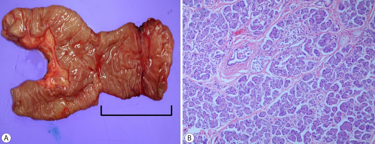
REFERENCES
3. Uppal K, Tubbs RS, Matusz P, Shaffer K, Loukas M. Meckelās diverticulum: a review. Clin Anat 2011;24:416ā422.   4. Park JJ, Wolff BG, Tollefson MK, Walsh EE, Larson DR. Meckel diverticulum: the Mayo Clinic experience with 1476 patients (1950-2002). Ann Surg 2005;241:529ā533.  5. Zarand A, Bajtai A, Baranyai Z, Dede K, Jakab F. Inflammation of ectopic pancreatic tissue in a Meckelās diverticulum causing acute abdominal symptoms: a case report and review of the literature. Int J Surg Pathol 2011;19:359ā363. 6. Lin S, Suhocki PV, Ludwig KA, Shetzline MA. Gastrointestinal bleeding in adult patients with Meckelās diverticulum: the role of technetium 99m pertechnetate scan. South Med J 2002;95:1338ā1341. 7. MĆ©ndez-GarcĆa C, SuĆ”rez-Grau JM, Rubio-Chaves C, MartĆn-Cartes JA, Docobo-DurĆ”ntez F, Padillo-Ruiz J. Surgical pathology associated with MeckelĀ“s diverticulum in a tertiary hospital: 12 year review. Rev Esp Enferm Dig 2011;103:250ā254.
|
|


