CME for
KSGE members
Choi and Moon: A rare case of intussusception in a patient with ulcerative colitis
Quiz
A 49-year-old female patient with ulcerative colitis (UC) who had opted out of treatment of her own accord visited the emergency room because of hematochezia. The day before visiting our hospital, the patient underwent sigmoidoscopy at a local hospital. Sigmoidoscopy revealed mucosal edema, erythema, and exudate in the rectum ( Fig. 1A). In addition, abdominopelvic computed tomography (CT) showed a mass like lesion at the hepatic flexure ( Fig. 1B). She was transferred to our hospital for further evaluation. Colonoscopy was performed the following day. A large circular mass that entirely blocked the lumen was observed at the hepatic flexure, and we performed a biopsy of the lesion to rule out colon cancer ( Fig. 1C). In addition, nodularity, erythema, friable mucosa, and geographical ulcerations were observed, extending from the distal sigmoid colon (20 cm from the anal verge) to the rectum. Pathological findings were unremarkable. On day 3, following hospitalization, follow-up abdominopelvic CT was performed to evaluate the bowel state, and the mass found previously at the hepatic flexure was no longer visible ( Fig. 1E). What is the most likely diagnosis?
Answer 
Pathologic examination showed acute and chronic inflammation with inflamed granulation tissue and the absence of cancerous lesions ( Fig. 2). The initial mass observed on colonoscopy was intussusception due to UC, which was reduced after colonoscopy. To manage the UC, we started treating the patient with mesalazine 1 g four times daily without steroids or any other UC medication from the admission date when she had previously stopped the treatment of her own accord. On day 3, following hospitalization, the abdominal pain and hematochezia were markedly reduced. On day 5, a diet was started, and on day 7, the patient was discharged with complete resolution of symptoms. There was no lesion in follow-up colonoscopy performed four months after discharge ( Fig. 3). Intussusception rarely causes acute abdominal pain in adults. It is even rarer in UC patients. In children, intussusception is usually caused by an idiopathic etiology; however, the condition is caused by an organic disease in the vast majority of adult cases (>90%). Small bowel intussusceptions are more common because of a benign cause, while the opposite is true for large bowel intussusceptions. 1 Most intussusceptions in adults are caused by neoplastic lesions, especially colonic intussusception, in which a tumor-related lesion serves as the lead point in >75% of cases. 2
The clinical manifestations of adult intussusception are variable, with abdominal pain, hematochezia, and vomiting being relatively common complaints. In some adults, no symptoms are observed. 3 An abdominal ultrasonographic examination or CT scan may be helpful in diagnosis, especially when a typical indicator suggestive of intussusception, such as the target sign or target mass, is noticed. Barium enema or air reduction is recommended for first-line management of pediatric patients. However, in adult intussusception, lead points are observed more often, and the incidence of malignancy is high. Therefore, preoperative barium or air reduction is not recommended. 4,5 Optimal management is through surgery, which is performed in almost all cases. 6
Fig.┬Ā1.
(A) Initial sigmoidoscopy revealed mucosal edema, erythema, and exudates. (B) Initial abdominopelvic computed tomography (CT) scan showed a mass like lesion at the hepatic flexure (arrow). (C, D) Follow-up colonoscopy revealed a mass-like lesion was observed at the hepatic flexure. (E) Follow-up abdominopelvic CT 3 days later showed the absence of a mass-like lesion at the hepatic flexure. 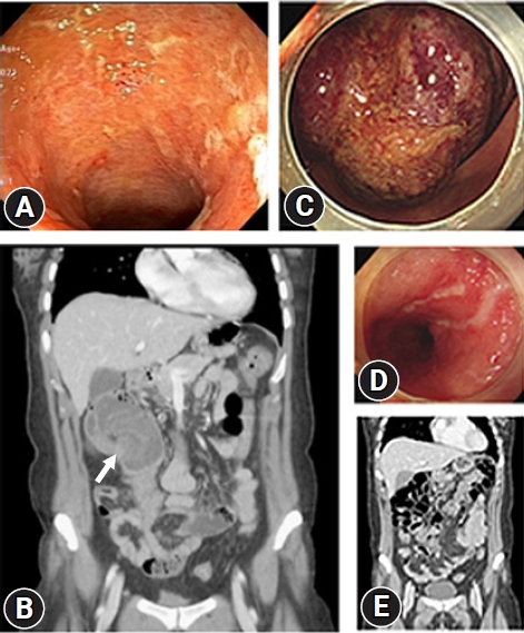
Fig.┬Ā2.
Histologically, the mass at the hepatic flexure was determined to be acute and chronic inflammation with inflamed granulation tissue (hematoxylin & eosin stain, ├Ś100). 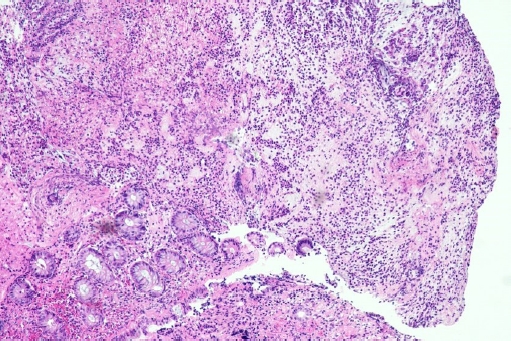
Fig.┬Ā3.
(A, B) Follow-up colonoscopy 4 months later revealed no lesion at the hepatic flexure and marked improvement of the inflamed mucosal lesions at the rectum. 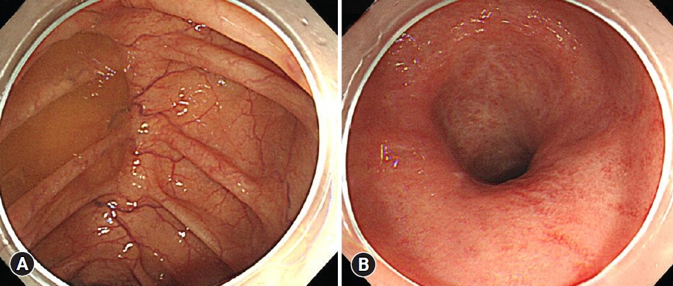
|
|


