CME for
KSGE members
Iraha, Kanemoto, and Hokama: All that elongates is not a polyp
Quiz
An 83-year-old woman with Sj├Čgren syndrome presented to the emergency unit with acute painless hematochezia. Her physical examination revealed unremarkable findings. Laboratory investigations showed a white blood cell count of 7,600/╬╝L, hemoglobin of 13.8 g/dL, and C-reactive protein of <0.1 mg/dL (reference range, <0.26 mg/dL). An enhanced abdominal computed tomography (CT) revealed an air bubble (arrow) and an extravasation (arrowhead) in the sigmoid colon ( Fig. 1). The subsequent colonoscopy ( Fig. 2) and narrow-band imaging (NBI) ( Fig. 3) revealed a hemorrhagic lesion. What is the most likely diagnosis?
Answer 
We suspected the air bubble revealed in the abdominal CT to be a colonic diverticulum before performing colonoscopy ( Fig. 1, arrow). NBI disclosed a normal mucosal pattern and concentric rings surrounding the polypoid lesion with an exposed vessel that made the diagnosis of an inverted colonic diverticulum (ICD) in the sigmoid colon ( Fig. 3). The exposed vessel was treated with endoscopic hemostatic clips following which hematochezia has not recurred. Although colonic diverticular disease is common, ICD has been rarely reported. 1-3 It often appears indistinguishable from the colon polyps. 2 The concentric rings, which are enhanced by NBI or indigo carmine dye, can help in diagnosing the ICD without maneuvers, such as the use of air insufflation, reverting the ICD with forceps, and the water jet deformation. 2,3 To the best of our knowledge, this is the first documentation of ICD associated with an exposed vessel that causes diverticular bleeding. This case emphasized the importance of recognition of ICD for endoscopists to avoid potentially risky procedures.
Fig.┬Ā1.
The abdominal computed tomography image showing an air bubble (arrow) and an extravasation of contrast (arrowhead) in the sigmoid colon. 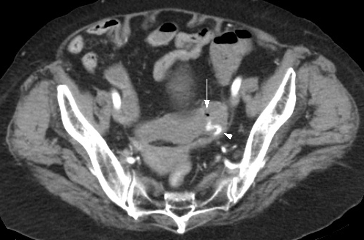
Fig.┬Ā2.
Colonoscopy revealing a polypoid lesion with an exposed vessel. 
Fig.┬Ā3.
Enhancement of the normal mucosal pattern and the concentric rings surrounding the polypoid lesion in narrow-band imaging. 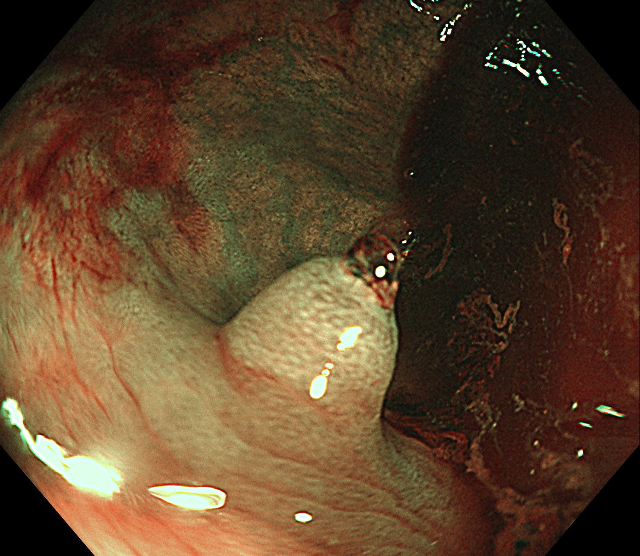
|
|


