Circumferential gastric wall thickening as initial presentation of relapsed plasma cell leukemia
Article information

Involvement of the gastrointestinal tract years after initial diagnosis of multiple myeloma (MM) is rare and often associated with a poor prognosis.1,2 In patients with newly diagnosed MM, the reported incidence of extramedullary MM (EMM) ranges from 0.5% to 4.8%, while in relapsed or refractory MM the reported incidence is 3.4% to 14%.3 The predominant sites of EMM involvement are the liver (28.8%), lymph nodes (23.1%), soft tissues of the neck (10%), breast (9%), adrenal glands (7.7%), and muscle (4.5%), with gastrointestinal tract reportedly involved in less than 5% of cases.4 There are some reports of recurrence as solitary masses or ulcers in the stomach.1,2,5,6 However, to the best of our knowledge, there have been no report of recurrence as diffuse thickening of the gastric wall. Here, we report the case of a patient with plasma cell leukemia (PCL) who relapsed with diffuse soft-tissue thickening of the gastric wall.
An eighty-year-old Japanese woman with lower back pain, which had been previously diagnosed as multiple pathological vertebral fractures, was referred to our hospital. Laboratory evaluation indicated a hemoglobin level of 10.1 g/dL, white blood cell count of 12.7×10⁹/L, and platelet count of 121×109/L. The peripheral blood smear showed 35% circulating abnormal plasmacytes; flow cytometry showed the cluster of differentiation (CD) 38+, CD138+, CD56+, and kappa>lambda. Other notable laboratory studies included serum creatinine of 0.75 mg/dL, calcium 8.7 mg/dL, lactate dehydrogenase (LDH) 263 U/L, beta-2 micro-globulin 6.76 mg/L, serum immunoglobulin (Ig) G 3,578 mg/dL, serum IgA 19 mg/dL, and serum IgM 7 mg/dL. Serum protein electrophoresis revealed IgG kappa with an M-spike (monoclonal protein). Bone marrow biopsy showed diffuse involvement with more than 80% plasma cells, and the karyotype was 46, XX. No cytogenetic abnormalities were detected. No EMM was found and the patient was diagnosed with International Staging System stage II IgG-kappa type PCL, an aggressive form of MM. The patient received induction chemotherapy with bortezomib and dexamethasone once a week for four weeks for five cycles, which resulted in a partial response. Monoclonal IgG levels decreased from 3,578 mg/dL before treatment to 653 mg/dL after induction therapy. The abnormal plasmacytes in the peripheral blood disappeared, however the LDH levels did not normalize. She presented with disease progression characterized by increased monoclonal IgG levels (1,012 mg/dL). Second-line chemotherapy with daratumumab, lenalidomide, and dexamethasone (DLd) was administered weekly in cycles 1 to 2, every two weeks in cycles 3 to 6, and every four weeks thereafter. Consequently, the disease stabilized with a very good partial response, and LDH levels normalized. After nine cycles of DLd therapy, the patient was switched to third-line chemotherapy (ixazomib combined with lenalidomide and dexamethasone) owing to grade four neutropenia. Two months after the initiation of third-line chemotherapy, the patient presented to the emergency department with abdominal swelling and poor oral intake. Laboratory studies disclosed the following: hemoglobin level 10.6 g/dL, white blood cell count 6,300/mm3 (abnormal plasmacytes, 28.5%), platelet count 80,000/mm3, serum creatinine 1.82 mg/dL, LDH 451 U/L, calcium 9.2 mg/dL, C-reactive protein 0.03 mg/L, serum ΙgG 1,311 mg/dL, serum IgA 5 mg/dL, and serum IgM 5 mg/dL. Abdominal computed tomography revealed circumferential soft-tissue thickening of the gastric wall and severe ascites (Fig. 1). Upper gastrointestinal endoscopy revealed gastric wall thickening and stiffening (Fig. 2). Additionally, the stomach was not fully distended despite air insufflation. Histological examination of the biopsy specimen revealed numerous clonal plasma cells infiltrating the gastric mucosal interstitium (Fig. 3). Immunohistochemical analysis revealed positivity for CD38 and an obvious predominance of kappa over lambda. Therefore, the patient was diagnosed with secondary EMM. The ascitic fluid contained numerous myeloma cells. Fluorescent in situ hybridization analysis of ascitic cells revealed TP53 deletion. She was intolerant of chemotherapy and died two weeks later because of MM.

Computed tomography image of the abdomen showing circumferential soft-tissue thickening of the gastric wall and a large amount of ascites.
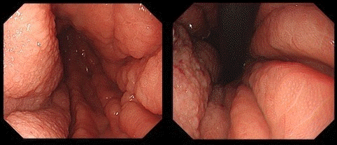
Endoscopic view showing thickening and stiffening of the entire stomach wall.
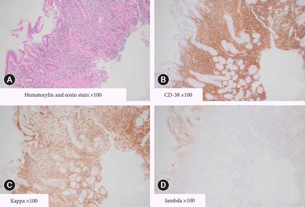
Histologic findings of the endoscopic biopsy specimen. (A) Diffuse dense infiltration of atypical plasma cells (hematoxylin and eosin stain, ×100). Immunohistochemical staining was positive for CD38 (B), and shows an obvious predominance of kappa (C) compared to lambda (D) (immunohistochemical stain, ×100).
There has been an overall increase in the incidence of EMM from 6.5% in 2005 to 23.7% in 2014.7 Furthermore, with the recent use of sensitive imaging techniques, EMM may be detected in up to 30% of MM patients during the course of their entire disease.8 Median time from diagnosis to occurrence of EMM has been observed to be 19 to 23 months.4 Both at diagnosis and at recurrence, EMM presentation was correlated with TP53 deletion in fluorescence in situ hybridization (FISH) analysis. In the present case, TP53 deletion was confirmed at the time of relapse.9 Gastrointestinal relapse in patients with MM who have been heavily pretreated, such as in our patient, is rare. In two large European studies, a total of 553 patients with MM, 87 of whom experienced extramedullary recurrences, none of which involved the gastrointestinal tract.5 To our knowledge, this is the first report of gastrointestinal relapse of PCL. The symptoms of EMM are typically associated with the lesion site. The clinical signs of gastrointestinal involvement include nonspecific gastrointestinal symptoms such as anorexia, weight loss, abdominal pain, vomiting, and gastrointestinal bleeding from ulcerative lesions. Gastrointestinal involvement may require to be considered as a differential diagnosis if gastrointestinal symptoms are present during the treatment of a patient with MM. In diffuse gastric diseases that extend throughout the stomach, thickening of the mucosal folds and hardening of the gastric wall have been observed. Differential diagnosis of diffuse gastric diseases, as based on sclerosis of the gastric wall and hypertrophy of the mucosal folds, may include scirrhous gastric carcinoma and malignant lymphoma, but is very rare in MM.10 In addition to more common causes (e.g., ulcer or mass), clinicians should consider gastrointestinal involvement in MM patients who present with thickening of the marginal soft tissue of the stomach wall when imaging studies are performed. This study was approved by the Institutional Ethics Board and was conducted in accordance with the principles of the Declaration of Helsinki. The patient provided informed consent for participation in the study and the publication of the report.
Notes
Conflicts of Interest
The authors have no potential conflicts of interest.
Funding
None.
Author Contributions
Conceptualization: YK; Supervision: MW; Investigation: YK, TK, AT; Writing–original draft: YK; Writing–review & editing: all authors.