Effective hemostasis under gel immersion endoscopy using inflated balloons on the tip of double balloon endoscope for active bleeding in the small intestine
Article information
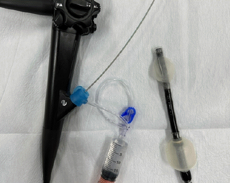
Securing an endoscopic visual field when identifying the source of gastrointestinal bleeding is difficult because the injected water easily mixes with blood and residue. Performing endoscopic procedures is challenging when the luminal contents are displaced by gas insufflation, which leads to significant discomfort. Gel immersion endoscopy (GIE) using an electrolyte-free gel (VISCOCLEAR®; Otsuka Pharmaceutical Factory) has been reported to be useful for securing the visual field during endoscopy for gastrointestinal bleeding.1,2 Particularly in the case of the small intestine, which is frequently filled with blood, GIE is effective for narrow lumens. This gel can easily replace the blood. Insufflation of the endoscope balloon avoids backflow of the gel, enabling it to push away blood and hold the intestinal wall, allowing the endoscope to stay at the targeted position. We also used a BioShield® irrigator (US Endoscopy), which allowed additional injection of the gel with a therapeutic device through the accessory channel. In addition, the use of a transparent cap makes it easier to concentrate the gel in front of the endoscope (Fig. 1). Herein, we report the successful treatment of a hemangioma with active bleeding in the small intestine under GIE using inflated balloons on the tip of a double balloon endoscope (DBE).
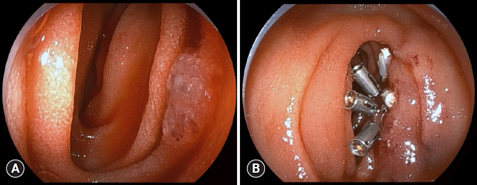
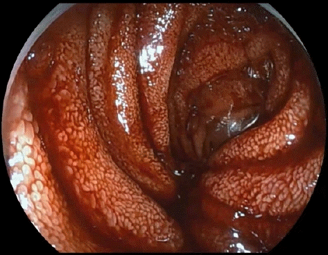
A 15-year-old boy had been suffering from melena for a month. Esophagogastroduodenoscopy and colonoscopy failed to reveal any signs of active bleeding. Capsule endoscopy was performed to check for small intestinal lesions and detected bloody intestinal fluid in the jejunum, followed by DBE, which revealed a large amount of fresh blood in the jejunum (Fig. 2). The cause of gastrointestinal bleeding was undetectable because the injected water was mixed with blood and the endoscope did not remain at the targeted position. However, by applying GIE using inflated balloons on the tip of the DBE, the visual field was secured, and the bleeding source was detected (Fig. 3, Supplementary Video 1). We immediately injected a hypertonic saline-epinephrine solution at the point of bleeding. Moderately active bleeding allowed us to secure the visual field. We were able to identify the lesion suspicious for a hemangioma as the source of bleeding, which was further performed with complete hemostasis using endoclips (SureClip®; Microtech) (Fig. 4). In this case, approximately 80 mL of gel was used. The patient was discharged without complications, and has not suffered from recurrence.

Gel immersion endoscopy using inflated balloons on the tip of double balloon endoscope secured the visual field, and the bleeding source was detected.

(A) After injection of hypertonic saline-epinephrine solution, the lesion suspicious of being hemangioma, which was the source of the bleeding, was visualized. (B) Hemostasis using endoclips was carried out.
In conclusion, GIE using inflated balloons on the tip of DBE is useful for precise treatment of active gastrointestinal bleeding, for it could facilitate endoscopic maneuvers and allow clear endoscopic view.
Supplementary Material
Supplementary Video 1.
Effective hemostasis under gel immersion endoscopy using inflated balloons on the tip of double balloon endoscope (https://doi.org/10.5946/ce.2023.146.v1).
Supplementary materials related to this article can be found online at https://doi.org/10.5946/ce.2023.146.
Notes
Conflicts of Interest
The authors have no potential conflicts of interest.
Funding
None.
Acknowledgments
We would like to thank members of the endoscopy room of Kansai Medical University Medical Center for their support.
Author Contributions
Conceptualization: SH; Investigation: SH, NS, HM, TM; Methodology: SH, NS, HM, TM, TY, MS; Project administration: all authors; Resources: SH, MS; Supervision: SH, MS; Validation: SH, MS; Visualization: SH; Writing–original draft: SH, MN; Writing–review and editing: SH, TY, MS, MN.


