Gastrointestinal Bleeding of Unknown Origin in Patients with Splenic Aneurysm
Article information
Quiz
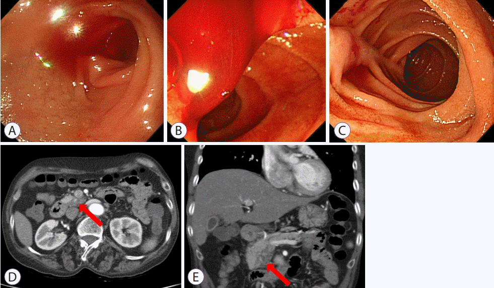
A 57-year-old woman was admitted to the hospital with melena and dyspnea. Her initial hemoglobin level was 6.5 g/dL. She had a history of acute pancreatitis with splenic pseudoaneurysm, which had been treated with angiographic coil embolization 1 year previously. After transfusion of several packs of red blood cells and tests for anemia, she underwent endoscopic examination. The colonoscopy findings were normal. Gastroscopic examination revealed blood in the second and third portions of the duodenum and suspicious active bleeding from the orifice of the ampulla of Vater (Fig. 1A-C). Because the bleeding focus was not clear, computer tomography (CT) scans and capsule endoscopy were performed. There was no evidence of bleeding in the small bowel during capsule endoscopy; however, CT scans revealed parenchymal swelling and an early enhancing lesion in the pancreatic uncinate process (Fig. 1D, E). What is the most likely diagnosis?
Answer
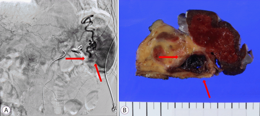
Given the focal early enhancing lesion in the pancreatic uncinate process, which was thought to reflect recent bleeding, hemosuccus pancreaticus was suspected. After CT scans, the patient underwent angiography but failed to receive additional embolization due to the previous coil (Fig. 2A). She finally underwent laparoscopic distal pancreatectomy because of the recurrent bleeding; the surgical specimen revealed a pseudoaneurysm at the splenic hilum (Fig. 2B).

(A) Celiac angiography shows a splenic artery pseudoaneurysm at the splenic hilum. (B) Surgical specimen demonstrates pseudoaneurysm with blood at splenic hilum.
Hemosuccus pancreaticus
Hemosuccus pancreaticus refers to bleeding from the pancreatic duct and is a rare cause of gastrointestinal bleeding [1]. It is most often found in patients with pancreatic tumors, pancreatitis, or pseudocysts. Bleeding occurs when surrounding vessels are eroded, and there is direct communication with the pancreatic duct. The most commonly involved vessel is the splenic artery [1]. It is often very aggressive and sometimes life-threatening.
Clinically, it must be suspected when upper gastrointestinal bleeding occurs and pancreatic injury is present. The best diagnostic modality is usually cross-sectional imaging, such as abdominal CT and/or magnetic resonance cholangiopancreatography. Endoscopic retrograde cholangiopancreatography may also be used for diagnosis [2-4].
Arteriography with coil embolization is usually the preferred treatment for acute bleeding control [3,4]. If bleeding persists or is massive, the treatment of choice is surgery with ligation of the bleeding vessel, which definitively prevents rebleeding [5,6].
Notes
Conflicts of Interest: The author has no potential conflicts of interest.

