Usefulness of the double-guidewire technique for endoscopic procedures in the field of biliary and pancreatic diseases
Article information

Abstract
The double-guidewire method has been increasingly used in endoscopic procedures for biliary and pancreatic diseases in recent years, including endoscopic retrograde cholangiopancreatography and endoscopic ultrasonography-related procedures. In addition, double-lumen catheters with uneven distal and proximal lumen openings have been introduced, making it possible to easily create a double-guidewire situation, and the usefulness of the double-guidewire technique using uneven double-lumen cannulas has been widely reported. Although the advantages of using two guidewires depend on the particular situation and the appropriate use of the two guidewires, deepening the knowledge of the double-guidewire method will contribute greatly to troubleshooting in daily practice. In this review, the usefulness of the double-guidewire technique is discussed with respect to two main areas: selective insertion of guidewires and devices and biliary cannulation.
INTRODUCTION
Endoscopic procedures for biliary and pancreatic disease, such as endoscopic retrograde cholangiopancreatography (ERCP) and endoscopic ultrasonography (EUS), cannot be performed without guidewires. There are currently three main guidewire thicknesses: 0.018, 0.025, and 0.035 inches. In addition to thickness, the properties of these guidewires could also greatly vary including material, tip shape, and length of the soft part.1 Currently, endoscopists can select an appropriate guidewire for individual cases from a vast list of guidewires. Usually, a single guidewire is required for a single procedure. For example, only one guidewire is required for wire-guided cannulation or the placement of plastic or metal stents. However, the usefulness of the double-guidewire technique (DGT) in various situations has been reported. The advantages of using two guidewires depend on the particular situation and their appropriate use. Hence, expanding our knowledge of the double-guidewire method will contribute to devising solutions in daily practice. Recently, a unique uneven double-lumen cannula (UDLC; PIOLAX, Tokyo, Japan) has been introduced. The UDLC is a double-lumen catheter with lumens measuring 0.025 and 0.035 inches in diameter. The orifice of each lumen is uneven, forming a channel at the tip of the UDLC (Fig. 1). UDLC allows the easy creation of a double-guidewire situation, and the usefulness of DGT using UDLC has been widely reported. In this review, the usefulness of DGT is examined with respect to two cardinal areas: its usefulness in the selective insertion of guidewires and devices, and its utility in biliary cannulation.
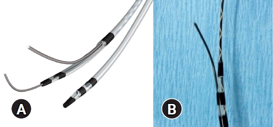
(A) Schema of the uneven double-lumen cannula (UDLC). The UDLC (PIOLAX, Tokyo, Japan) is a double-lumen catheter with lumens measuring 0.025 (distal) and 0.035 (proximal) inches in diameter. The orifice of each lumen is uneven, creating a channel at the tip. (B) A guidewire can be manipulated from the proximal lumen of the UDLC.
USEFULNESS OF THE DOUBLE-GUIDEWIRE METHOD IN THE SELECTIVE INSERTION OF GUIDEWIRES AND DEVICES
There are two situations in which the DGT is useful for guidewire manipulation and device insertion. The first is to demonstrate the usefulness of guidewires with different properties for selective insertion. The other is to improve the straightness of device insertion using two guidewires and achieve security even if one guidewire comes loose. DGT has been shown to be useful in various ERCP-related transpapillary procedures and EUS-guided drainage.
Usefulness in ERCP-related transpapillary procedures
There are several different types of benign bile duct stenoses. Among these, postoperative bile duct stenosis is non-physiological and associated with a high degree of narrowing. Therefore, it is difficult to insert a guidewire beyond the stenosis site. In a report on the usefulness of UDLC, DGT using two different types of guidewires, hydrophilic (Radifocus; Terumo, Tokyo, Japan) and stiff (VisiGlide2; Olympus, Tokyo, Japan) guidewires, led to successful drainage beyond postoperative bile duct stenosis.2 In this case, a hydrophilic guidewire, which had excellent penetration, was used to successfully penetrate the stenosis; however, stent insertion was not possible because of insufficient guidewire stiffness. Therefore, UDLC was used to insert a stiff guidewire beyond the stenosis by using the proximal lumen. Because of the successful insertion of the two guidewires, the stenosis was slightly relieved and the device could be inserted beyond the stenosis. This case illustrates the importance of understanding the characteristics of each guidewire and using them appropriately in each situation.
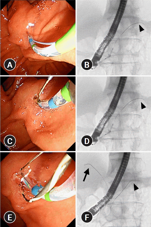
In addition, DGT using UDLC has been reported to be useful for stent-in-stent implantation in hilar bile duct stenosis.3 One of the absolute prerequisites for successful stent-in-stent placement of metallic stents (MSs) is that the guidewire for the second MS must be inserted into the drainage area through the mesh of the first MS. As the MS used for stent-in-stent placement is uncovered, the guidewire can easily cross the mesh on the papillary side instead of the hilar side. If the guidewire does not pass through the hilar mesh of the first MS, insertion of the second MS will be unsuccessful, leading to deformation of the first MS. However, it is impossible to determine whether the guidewire has passed through the mesh of the first MS on the fluoroscopy screen. DGT using UDLC can solve this problem. The UDLC is inserted into the hilar region using the same guidewire that was used to place the first MS, and the guidewire for the second MS is inserted through the proximal lumen into the drainage branch ready for stent-in-stent placement (Fig. 2).
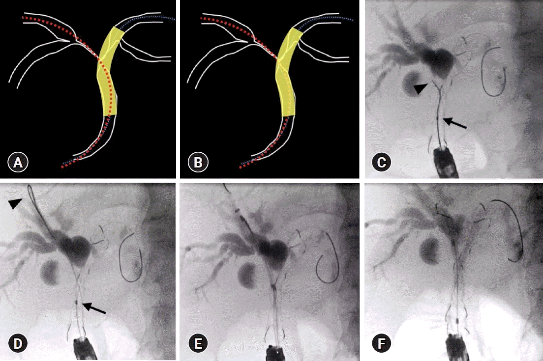
Double-guidewire method with uneven double-lumen cannula for the placement of a self-expandable metal stent (SMS) using the stent-in-stent (SIS) method. (A) Guidewire situation where the SIS method was unsuccessful. The guidewire for the second stent insertion was not passed through the mesh of the first stent. (B) Guidewire situation where SIS was successful. The guidewire for the second stent insertion is passed through the mesh of the first stent. (C) The uneven double-lumen cannula was inserted into the stent using the guidewire used for the first stent placement (arrow), and the guidewire tip (arrowhead) from the proximal lumen passed through the stent mesh. (D) The UDLC was inserted into the stent using the same guidewire used for the first stent placement (arrow), and the guidewire tip (arrowhead) was guided from the proximal lumen into the drainage area. (E) The second stent delivery was through the mesh of the first stent. (F) Successful SMS placement using the SIS method.
Patients with hilar cholangiocarcinoma may have multiple plastic stents inserted, instead of metal stents. Therefore, multiple guidewire insertion techniques are very important when inserting two or more, especially up to three plastic stents. In such situations, a swing catheter or sphincterotome is useful for selective guidewire manipulation of the bile duct. These methods are also broadly included in the DGTs.
Usefulness of the DGT in EUS-guided cyst drainage
EUS-guided cyst drainage is performed for infected pancreatic cysts and walled-off necrosis (WON).4,5 In recent years, use of lumen-apposing metal stents has been widely reported.6-9 However, there are some institutions and regions where lumen-apposing metal stents are not available, and plastic stent placement still plays an important role in the treatment of infected pancreatic cysts and WON. WON cases require removal of internal necrotic tissue during endoscopic or surgical necrosectomy. It has also been reported that intravenous treatment using a nasal drainage route prevents necrosectomy,10 and internal and external fistula treatment with nasal drainage is often used in addition to only one procedure of the pancreatic stent-assisted method (PS) for drainage treatment. In addition, a step-up approach has been established as a treatment strategy for WON,11 and drainage with multiple stents is recommended as a useful method.12-16 In multiple stenting, the site of stent placement is important and multiple guidewire manipulations are required.
Itoi et al.17 reported the usefulness of creating a double-guidewire situation and then using two guidewires for the simultaneous placement of a plastic stent and nasocystic drainage tube. They reported a procedure success rate of 92% (12/13), a clinical success rate of 100% (13/13), no procedure-related complications, and no recurrence within a mean observation period of 785 days. Seewald et al.18 developed a kit that allows simultaneous placement of two guidewires and reported its usefulness. UDLC enables easy double-guidewire insertion and is expected to be useful for the double placement of a plastic stent and nasocystic drainage tube (Fig. 3). The DGT is useful for EUS-guided cyst drainage.
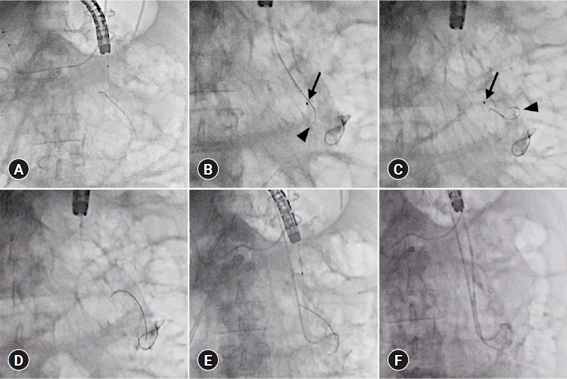
Double-guidewire method using the uneven double-lumen cannula for endoscopic ultrasonography cyst drainage. (A) One guidewire was inserted into the lumen of the walled-off necrosis (WON). (B) The uneven double-lumen cannula (arrow) was inserted into the WON, and the guidewire (arrowhead) was manipulated from the proximal lumen. (C) The UDLC (arrow) was inserted into the WON, and the guidewire (arrowhead) was inserted deep into the WON. (D) Double-guidewire situation. (E) First, a plastic stent was inserted. (F) Thereafter, a nasocystic drainage tube was inserted.
Usefulness of the DGT in EUS-guided biliary drainage
EUS-guided biliary drainage consists of the following steps: trans-gastrointestinal bile duct puncture, guidewire placement, puncture site dilation, and stent placement. However, these steps are difficult in cases where scope stability is impaired.19,20 Even if the guidewire is placed in the drainage area, there is a possibility that the guidewire will pull out if the scope position is difficult to maintain.
Shiomi et al.21 reported that increasing the number of guidewires from one to two can improve the scope stability, and Mandai et al.22 reported that stability with a double-guidewire was useful during EUS-guided antegrade stenting.
Kawakami et al.23 reported that guidewire manipulation from the proximal lumen using UDLC was useful for manipulating the guidewire to the hilar side of the liver in EUS-hepaticogastrostomy, which is occasionally difficult. Similarly, Ishiwatari et al.24 reported the usefulness of UDLC for access from the left bile duct to the right bile duct during EUS-hepaticogastrostomy in patients with malignant hilar biliary obstruction. Nakai et al.25 reported on DGT using UDLC for EUS-guided drainage at their institution and listed multiple drainage placements, scope stabilization, device insertion, safety guidewire, and antegrade stone removal as the main reasons for using DGT. They reported that the success rate of UDLC insertion was 100%, that of the preplanned technique was 92.7%, and that there were no complications related to DGT.
USEFULNESS OF THE DOUBLE-GUIDEWIRE METHOD FOR BILIARY CANNULATION
Biliary cannulation is a basic but challenging ERCP-related procedure. Without successful biliary cannulation, the planned procedure cannot be performed. The pancreatic guidewire (PGW) placement method, which is a double-guidewire method, and the uneven method have been reported to be useful in cases of difficult biliary cannulation. Both methods are DGTs that use a pancreatic ductal guidewire, but they have different characteristics.
The PGW method
In the PGW method, the papilla is fixed by a PGW and biliary cannulation is attempted using another guidewire (Fig. 4). Dumonceau et al.26 first reported the PGW method in 1998 as an effective method for biliary cannulation in postoperative gastric reconstruction after Billroth I surgery. Since then, the PGW method has been widely used in clinical practice, not only for postoperative anatomy but also for cases of difficult biliary cannulation with a normal anatomy.
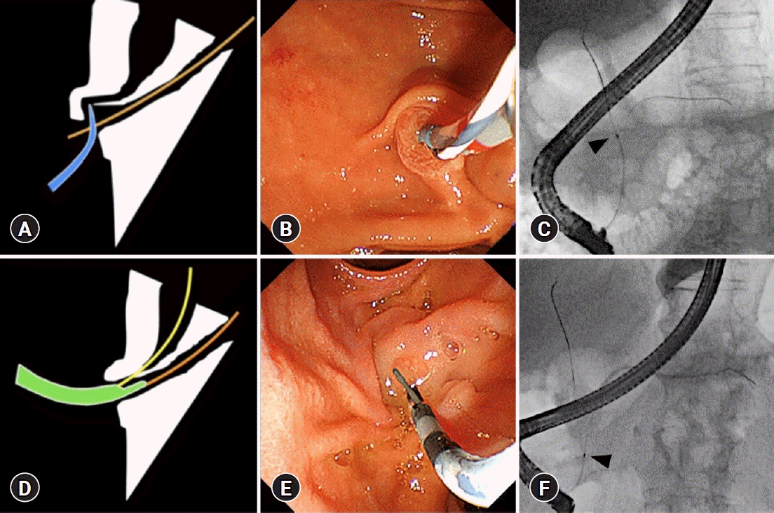
Double-guidewire method using the uneven double-lumen cannula (UDLC) for biliary cannulation. (A) Schema of the pancreatic duct guidewire method. (B) The papilla was fixed with a guidewire and a cannula was used for biliary cannulation. (C) A cannula (arrowhead) was inserted into the bile duct. (D) Schema of the uneven method. (E) The papilla was fixed with the UDLC itself, and biliary cannulation was attempted with a guidewire from the proximal lumen. (F) The papilla was fixed by the UDLC (arrowhead), and a guidewire from the proximal lumen was inserted into the bile duct.
The usefulness of the PGW method has been evaluated, but the results vary between reports (Table 1).27-36 The reasons for this variation include the fact that the studies were conducted with diverse definitions of “difficult biliary cannulation” and that the technique compared with the PGW method differed between the studies. As shown in Table 1, the recognized time for “difficult biliary cannulation” varied from 5 to 15 minutes after the start of the test. Two main techniques were compared: normal techniques and special techniques, such as PS and transpancreatic sphincterotomy (TPS). TPS is a useful biliary cannulation and papillotomy technique using a pancreatic duct guidewire (Fig. 5). TPS can also be considered as belonging to the DGT in a broader sense.

Outcomes of the pancreatic guidewire method
Transpancreatic sphincterotomy. (A, B) The papilla was fixed with a pancreatic guidewire (arrowhead) and a sphincterotome was inserted using the pancreatic guidewire. (C, D) Sphincterotomy was performed with the pancreatic duct guidewire in place (arrowhead). (E, F) Successful wire-guided biliary cannulation was achieved (arrow) using the sphincterotome with the pancreatic duct guidewire in place (arrowhead).
Compared with the conventional method, most reports have shown a higher success rate of biliary cannulation using the PGW method. A recent randomized controlled trial (RCT) by Laquiere et al.32 reported that early use of the PGW method improved the rate of biliary cannulation within 10 minutes. However, there are many reports of lower success rates for biliary cannulation using PGW compared with PS and TPS. However, although post-ERCP pancreatitis (PEP) is occasionally fatal,37 the impact of the PGW method on the incidence of PEP tended to be similar in many reports, and the use of the PGW method tended to increase the incidence of PEP.
There have been three meta-analyses on PGW methods.38-40 In a meta-analysis of seven RCTs by Tse et al.,40 the PGW method was reported to increase the risk of PEP without any superiority in achieving biliary cannulation compared with other techniques. Another meta-analysis of four RCTs by Guzman-Calderon et al.,39 comparing the PGW method and TPS, showed that the PGW method had a lower success rate in biliary cannulation and a higher incidence of PEP than TPS. In one of the four RCTs in this meta-analysis, the incidence of PEP in the PGW group was 38.2%, and the incidence of complication was notably high (83.9%).36 In another RCT, the incidence of PEP in the PGW group was high (47.4%).41 A recent meta-analysis reported that early needle-knife techniques and TPS were superior to other interventions in decreasing PEP rates, and should be considered in patients with difficult biliary cannulation.38 According to the European Society of Gastrointestinal Endoscopy guidelines for ERCP-related adverse events, a pancreatic duct stent is recommended to prevent PEP whenever the PGW method is used.42 Wang et al.43 in 2021 reported that the combined use of indomethacin and pancreatic ductal stents reduced the incidence of PEP compared with the use of indomethacin alone in the PGW group. In summary, the PGW method is undoubtedly a useful option for patients with difficult biliary cannulation. However, the risk of PEP requires careful consideration.
The uneven method
The uneven method is a variation of the PGW method that uses UDLC. The uneven method is performed as follows: (1) insertion of the UDLC tip into the papilla with PGW assistance and (2) wire-guided biliary cannulation using the proximal lumen of the UDLC (Fig. 4).44
Linearization of the common channel and fixation of the papilla produced by UDLC makes it easy to adjust the axis of the catheter in the direction of the bile duct, leading to successful biliary cannulation. The uneven and PGW methods are similar in that both are methods of biliary cannulation assisted by fixation of the papilla; however, while the PGW method uses a guidewire, the uneven method uses a catheter to fix the papilla. Therefore, the uneven method is expected to provide stronger papillary fixation and straightening of the bile ducts than the PGW method. In addition, the uneven method has the advantage of eliminating this problem, whereas in the PGW method, the guidewire may interfere with the approach of the catheter tip to the bile duct opening.
A novel method of biliary cannulation has been reported, with a concept that differs from DGT, in which a UDLC is inserted into the papilla without PGW and biliary cannulation is performed using the proximal lumen of the UDLC.45
In addition, this method has also been reported to be useful for balloon enteroscopy-assisted ERCP in patients with a surgically altered gastrointestinal anatomy.46 In a comparative study of the two techniques in 86 cases of balloon enteroscopy-assisted ERCP, the success rate of biliary cannulation tended to be higher in the uneven method (83.3%) than in the PGW method (59%). The cannulation time was also significantly shorter in the uneven method. PEP occurred in 13.6% of the PGW group, but not in the uneven method.47 A prospective study is currently underway, and it is expected that this uneven method may be a useful option for patients with difficult biliary cannulation.
CONCLUSIONS
In this review, the usefulness of DGT was outlined with respect to two main issues: its usefulness in the selective insertion of guidewires and devices, and its utility in biliary cannulation. The DGT contributes to endoscopic procedures in various situations. Currently, UDLC is only available in Japan, but other devices are being developed using the same concept. To maximize the merits of using two guidewires, it is important to understand the characteristics of each guidewire and to select the appropriate type for each case.
Notes
Conflicts of Interest
The authors have no potential conflicts of interest.
Funding
None.
Author Contributions
Supervision: MK; Writing–original draft: MT; Writing–review & editing: MT, MK.