Skip Navigation
Skip to contents

Clin Endosc : Clinical Endoscopy

OPEN ACCESS

Articles

Page Path
HOME > Clin Endosc > Volume 46(2); 2013 > Article
Original Article Diagnostic Accuracy and Interobserver Agreement in Predicting the Submucosal Invasion of Colorectal Tumors Using Gross Findings, Pit Patterns, and Microvasculatures
Hye Jung Choi, Bo-In Lee, Hwang Choi, Kyu Yong Choi, Sang-Woo Kim, Joo Yong Song, Jeong Seon Ji, Byung-Wook Kim
Clinical Endoscopy 2013;46(2):168-171.
DOI: https://doi.org/10.5946/ce.2013.46.2.168
Published online: March 31, 2013

Division of Gastroenterology, Department of Internal Medicine, The Catholic University of Korea College of Medicine, Seoul, Korea.

Correspondence: Bo-In Lee. Division of Gastroenterology, Department of Internal Medicine, Incheon St. Mary's Hospital, The Catholic University of Korea, 56 Dongsu-ro, Bupyeong-gu, Incheon 403-720, Korea. Tel: +82-32-280-5052, Fax: +82-32-280-5987, gidoc4u@catholic.ac.kr
• Received: May 28, 2012   • Revised: October 24, 2012   • Accepted: October 24, 2012

Copyright © 2013 Korean Society of Gastrointestinal Endoscopy

This is an Open Access article distributed under the terms of the Creative Commons Attribution Non-Commercial License (http://creativecommons.org/licenses/by-nc/3.0/) which permits unrestricted non-commercial use, distribution, and reproduction in any medium, provided the original work is properly cited.

  • 7,722 Views
  • 59 Download
  • 7 Crossref
  • 6 Scopus
prev next
  • Background/Aims
    Depth of invasion is one of the most important factors for establishing treatment strategy for colorectal tumors.
  • Methods
    Three blinded experts reviewed electronic photos and video clips of 33 early colorectal cancer-like lesions. They estimated the depth of invasion based on conventional white light endoscopy (CWE), magnifying chromoendoscopy (MCE), and magnifying narrow band imaging endoscopy (MNE).
  • Results
    The lesions included nine mucosal low-grade neoplasias, 16 mucosal high grade neoplasias, and eight carcinomas with invasion to the submucosal layer or beyond. The diagnostic accuracy for submucosal invasion by CWE ranged from 67% to 82%, while those by MCE and MNE ranged from 85% to 88% and 85% to 88%, respectively. The diagnostic accuracy significantly differed between CWE and MCE (p=0.034) and between CWE and MNE (p=0.039). The kappa values for CWE, MCE, and MNE among the endoscopists were 0.564, 0.673, and 0.673, respectively.
  • Conclusions
    The estimation of submucosal invasion for early colorectal cancer-like lesions based on MCE or MNE is more accurate than CWE. MCE and MNE were demonstrated to have substantial agreement for predicting submucosal invasion.
Estimation of invasion depth is one of the most important steps for establishing therapeutic plans for colorectal tumors. Without deep submucosal invasion, lymph node metastasis rarely occurs in colorectal tumors,1-3 and those tumors can be treated with local excision such as endoscopic mucosal resection (EMR) or endoscopic submucosal dissection (ESD).
The depth of invasion of colorectal tumors can be estimated by using the gross findings of conventional white light endoscopy (CWE), pit patterns identified by magnifying chromoendoscopy (MCE),4,5 and the surface microvascular patterns identified by magnifying narrow band imaging endoscopy (MNE).6-8 However, their accuracy and interobserver agreement were rarely reported.
Thus, this study was performed to evaluate the accuracy and interobserver agreement of expert endoscopists for predicting the depth of invasion using CWE, MCE, and MNE.
Subjects
All of the CWE, MCE, and MNE electronic photos and video clips of the cases were recorded with magnifying colonoscopes (CF-H260AZL; Olympus Co., Tokyo, Japan) by an expert endoscopist (B.I.L) from November 2009 to October 2010. Thirty-three colorectal tumors were included and all the tumors had more than one of gross features suggesting early colorectal cancer (Tis or T1); which are hardness, surrounding white spots, depression, surface nodularity, full expansion, deformed adjacent wall, mucosal friability, convergence of mucosal folds, erosion, or ulceration.9,10 Grossly obvious advanced cancers with luminal obstruction or large ulceration were excluded.
All of the photos and video clips were taken from as many angles as possible after washing the lesions with simethicone-dissolved water. The CWE was recorded first, followed by MNE and MCE. Observation with magnifying endoscopy was performed more carefully for depressed or erosive areas. MCE was performed with 0.4% indigo carmine and 0.05% crystal violet in sequence. When crystal violet was applied, observation was not made until the dye had sufficiently stained the tumor.
All the final histopathologic results of the resected specimens were reclassified according to the Revised Vienna Classification of Gastrointestinal Neoplasia (Table 1).11
Estimation of invasion depth
Three expert endoscopists (S.W.K, H.C, and K.Y.C) independently reviewed the electronic photos and video clips of the cases. The endoscopists had experiences of more than 10,000 colonoscopy cases and had used magnifying endoscopy for several years.
Kudo's classification was used for pit pattern analysis.12 Microvascular patterns were categorized according to the Showa Classification.7,13 Unlike pit pattern, classifications for microvascular pattern had not been standardized at the time of the study,14 and the Showa Classification was therefore reviewed by the endoscopists before the estimation of invasion depth.
Only the information on sex and age of the patients was provided to the endoscopists. The endoscopists reviewed the CWE, the MCE, and the MNE images in sequence.
Based on the CWE, the endoscopists evaluated gross findings suggestive of submucosal invasion (depression, full expansion, wall deformity, spontaneous bleeding, mucosal convergence, erosion, or ulcer)5,15 and the depth of invasion (mucosa vs. submucosa or beyond). The estimations were made again based on the MCE, and then on the MNE. A "VN pattern" on the MCE12 and a "sparse pattern" on the MNE7,13 were considered suggestive of submucosal invasion (Fig. 1).
The study protocol was approved by the Institutional Review Board (OC11RIMI0011).
Statistical analysis
The SAS system version 9.1 (SAS Institute Inc., Cary, NC, USA) was used for statistical analysis. Differences of diagnostic accuracy for submucosal invasion among the endoscopists and among the methods of endoscopy were evaluated by Generalized Estimating Equations. The p-values were corrected by Bonferroni's method. The interobserver agreement among the endoscopists was evaluated by Fleiss' kappa.
Baseline characteristics
The median diameter of the lesions was 24 mm (range, 10 to 61). The lesions included 13 protruded type, one flat type, two depressed type, and 17 laterally spreading tumors. The tumors were treated primarily by EMR (11), ESD (17), and surgery (5). One of the patients who had undergone EMR received additional surgery later due to invasion to the deep submucosal layer. The histopathologic results were as follows: mucosal low-grade neoplasia (low grade adenoma/dysplasia) in nine patients; mucosal high grade neoplasia (high grade adenoma/dysplasia, noninvasive carcinoma, and intramucosal carcinoma) in 16 patients; and submucosal or further invasion by carcinoma) in eight patients.
Diagnostic accuracy for submucosal invasion
Among the endoscopists, the diagnostic accuracy for submucosal invasion using gross findings of CWE ranged from 67% to 82%, while those using pit patterns identified by MCE and microvasculature patterns identified by MNE ranged from 85% to 88% and 85% to 88%, respectively (Fig. 2).
There was no significant difference in diagnostic accuracy among the endoscopists (p=0.257). However, the diagnostic accuracy significantly differed among the methods of endoscopy (p=0.040). The interaction between the endoscopists and the methods of endoscopy was not significant (p=0.086).
The diagnostic accuracy significantly differed between CWE and MCE (p=0.034, Bonferroni's correction) and between CWE and MNE (p=0.039, Bonferroni's correction). However, the diagnostic accuracy did not significantly differ between MCE and MNE (p>0.05).
Interobserver agreement
The kappa value for CWE among the three endoscopists was 0.564. Kappa values for MCE and MNE were 0.673 and 0.673, respectively.
"Early colorectal cancer" is defined by Japanese doctors as colorectal cancer limited to the mucosa or invading only the submucosa, regardless of the presence or absence of lymph node metastases.16 However, in this study, we evaluated the diagnostic accuracy of submucosal invasion for "early colorectal cancer-like lesions" instead of "pathologically confirmed early colorectal cancers." Only the gross features suggesting early colorectal cancer9,10 were used for the selection criteria.
The evaluation of early colorectal cancer-like lesions can be more practical than the evaluation of confirmed early colorectal cancers; this is because, first, it is not always easy to achieve endoscopic differentiate between a carcinoma and an adenoma or even between early cancer and advanced cancer. In other words, when an early colorectal cancer-like tumor is detected during a colonoscopy, we cannot confirm whether the lesion is an early colorectal cancer until the final pathology report of the resected specimen has been obtained. The role of forceps biopsy is also limited since the histopathology of endoscopic forceps biopsy disagrees with that of a resected specimen in 40% of cases.17
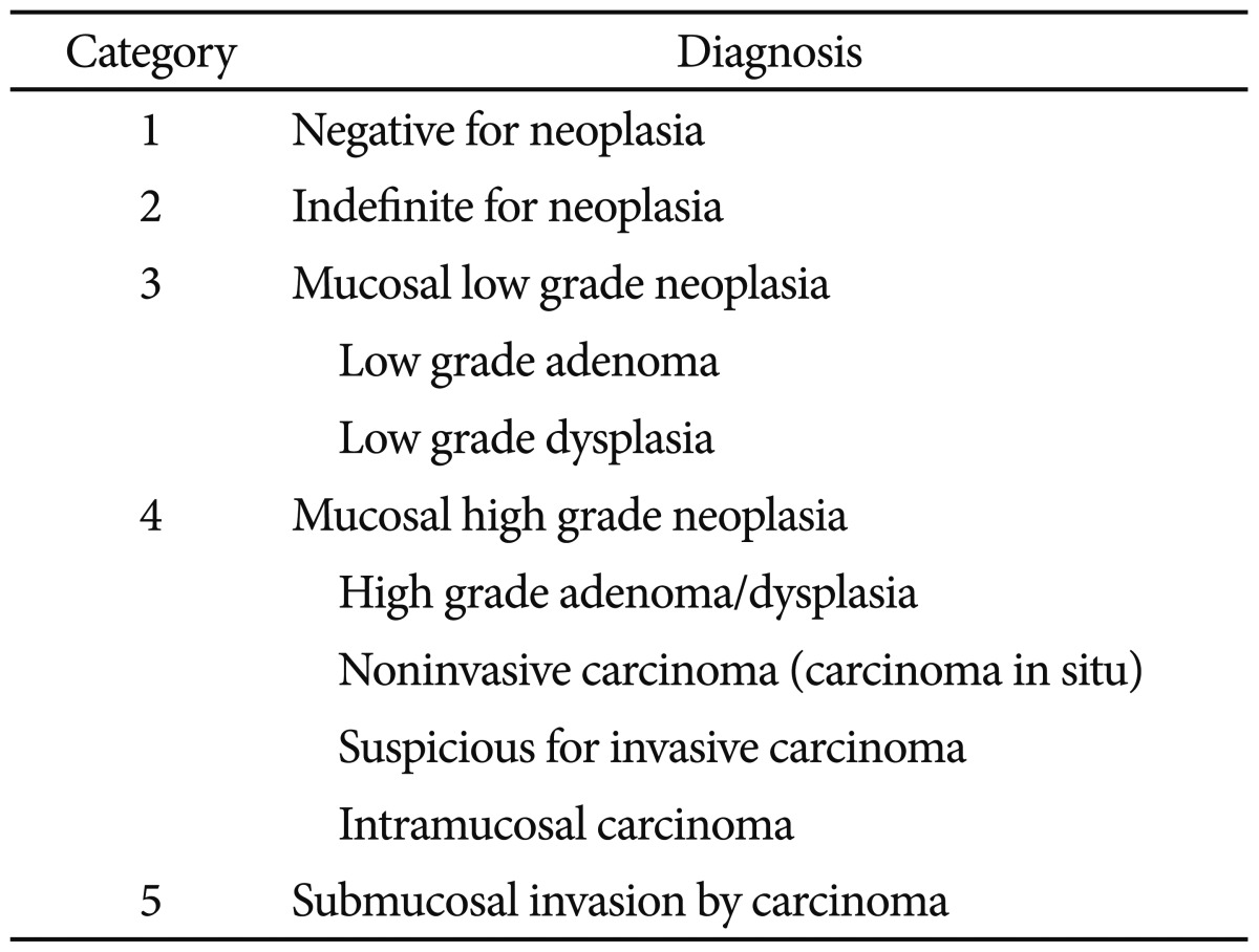
Second, when establishing therapeutic plans, it may be more important to determine whether the tumor is limited to the mucosal layer rather than whether the tumor is a carcinoma or an adenoma. Most mucosal neoplasms (revised Vienna classification categories 3 and 4:11 low grade adenoma/dysplasia, high grade adenoma/dysplasia, noninvasive carcinoma, and intramucosal carcinoma) can be treated by local excision (EMR or ESD) because lymph node metastasis is rarely reported without deep submucosal invasion.
The diagnostic accuracy for submucosal invasion was significantly improved by pit pattern analysis with MCE in this study. Moreover, the interobserver agreement of MCE was substantial, while the interobserver agreement of CWE was moderate. As such, MCE can estimate the depth of invasion for early colorectal cancer-like lesions more accurately and more objectively.
The diagnostic accuracy for submucosal invasion also significantly differed between CWE and MNE. However, the diagnostic accuracy did not differ between MCE and MNE. The estimation of submucosal invasion using MNE might be limited since the irregular microvascular pattern by the Showa classification could be interpreted in different ways. We considered an "irregular microvascular pattern" identified using MNE as a mucosal cancer without submucosal invasion. However, according to other studies,13,14 more than half of colorectal tumors with an irregular microvascular pattern showed deep submucosal invasion. In addition, microvascular classifications of colorectal tumors based on MNE had not been unified at the time of the study,14 and all of the expert endoscopists in this study were more familiar with pit pattern analysis using MCE than microvascular pattern analysis using MNE.
It is possible that the results would have been different if the endoscopists viewed the MNE images before the MCE images. In practice, MNE is performed before MCE because the applied dye used for chromoscopy interferes with MNE. However, in this study, estimation was performed earlier by MCE than by MNE because we set out to identify any additional effects of MNE on MCE for predicting submucosal invasion.
After the depth of invasion was estimated, we examined the cases in which the results of MCE and MNE did not agree among the endoscopists. The reasons for disagreement were mixed pit/microvascular patterns and bad images due to bleeding, exudation, or insufficient staining.
In conclusion, the estimation of submucosal invasion based on MCE or MNE is more accurate and more helpful for establishing a treatment strategy for colorectal tumors than the estimation based on CWE. MCE and MNE were demonstrated to have substantial agreement for estimating the depth of invasion.
The study consultation with Catholic Research Coordinating center was supported by the Korea Health 21 R&D Project (A070001), Ministry of Health & Welfare, Republic of Korea.
  • 1. Kitajima K, Fujimori T, Fujii S, et al. Correlations between lymph node metastasis and depth of submucosal invasion in submucosal invasive colorectal carcinoma: a Japanese collaborative study. J Gastroenterol 2004;39:534–543.ArticlePubMed
  • 2. Ueno H, Mochizuki H, Hashiguchi Y, et al. Risk factors for an adverse outcome in early invasive colorectal carcinoma. Gastroenterology 2004;127:385–394.ArticlePubMed
  • 3. Okabe S, Shia J, Nash G, et al. Lymph node metastasis in T1 adenocarcinoma of the colon and rectum. J Gastrointest Surg 2004;8:1032–1039.ArticlePubMed
  • 4. Kudo S, Rubio CA, Teixeira CR, Kashida H, Kogure E. Pit pattern in colorectal neoplasia: endoscopic magnifying view. Endoscopy 2001;33:367–373.ArticlePubMed
  • 5. Ikehara H, Saito Y, Matsuda T, Uraoka T, Murakami Y. Diagnosis of depth of invasion for early colorectal cancer using magnifying colonoscopy. J Gastroenterol Hepatol 2010;25:905–912.ArticlePubMed
  • 6. Kanao H, Tanaka S, Oka S, Hirata M, Yoshida S, Chayama K. Narrow-band imaging magnification predicts the histology and invasion depth of colorectal tumors. Gastrointest Endosc 2009;69(3 Pt 2):631–636.ArticlePubMed
  • 7. Wada Y, Kudo SE, Kashida H, et al. Diagnosis of colorectal lesions with the magnifying narrow-band imaging system. Gastrointest Endosc 2009;70:522–531.ArticlePubMed
  • 8. Ikematsu H, Matsuda T, Emura F, et al. Efficacy of capillary pattern type IIIA/IIIB by magnifying narrow band imaging for estimating depth of invasion of early colorectal neoplasms. BMC Gastroenterol 2010;10:33.ArticlePubMedPMCPDF
  • 9. Mainprize KS, Mortensen NJ, Warren BF. Early colorectal cancer: recognition, classification and treatment. Br J Surg 1998;85:469–476.ArticlePubMed
  • 10. Kudo S, Kashida H, Nakajima T, Tamura S, Nakajo K. Endoscopic diagnosis and treatment of early colorectal cancer. World J Surg 1997;21:694–701.ArticlePubMed
  • 11. Dixon MF. Gastrointestinal epithelial neoplasia: Vienna revisited. Gut 2002;51:130–131.ArticlePubMedPMC
  • 12. Kudo S, Tamura S, Nakajima T, Yamano H, Kusaka H, Watanabe H. Diagnosis of colorectal tumorous lesions by magnifying endoscopy. Gastrointest Endosc 1996;44:8–14.ArticlePubMed
  • 13. Wada Y, Kudo SE, Misawa M, Ikehara N, Hamatani S. Vascular pattern classification of colorectal lesions with narrow band imaging magnifying endoscopy. Dig Endosc 2011;23(Suppl 1):106–111.ArticlePubMed
  • 14. Tanaka S, Sano Y. Aim to unify the narrow band imaging (NBI) magnifying classification for colorectal tumors: current status in Japan from a summary of the consensus symposium in the 79th Annual Meeting of the Japan Gastroenterological Endoscopy Society. Dig Endosc 2011;23(Suppl 1):131–139.ArticlePubMed
  • 15. Ji JS, Choi H, Choi KY, et al. Interobserver agreement among experts in determining the depth of invasion of early colorectal carcinoma. Korean J Gastrointest Endosc 2005;31:39–43.Article
  • 16. Kashida H, Kudo SE. Early colorectal cancer: concept, diagnosis, and management. Int J Clin Oncol 2006;11:1–8.Article
  • 17. Sung HY, Cheung DY, Cho SH, et al. Polyps in the gastrointestinal tract: discrepancy between endoscopic forceps biopsies and resected specimens. Eur J Gastroenterol Hepatol 2009;21:190–195.ArticlePubMed
Fig. 1
Magnifying endoscopy findings suggesting submucosal invasion. (A) The chromoendoscopy VN pattern. (B) The narrow band imaging endoscopy sparse pattern.
ce-46-168-g001.jpg
Fig. 2
The diagnostic accuracy of submucosal invasion for each endoscopist according to the method of endoscopy. NBI, narrow band imaging.
ce-46-168-g002.jpg
Table 1
The Revised Vienna Classification of Gastrointestinal Epithelial Neoplasia
ce-46-168-i001.jpg

Figure & Data

REFERENCES

    Citations

    Citations to this article as recorded by  
    • Underwater Versus Conventional Endoscopic Mucosal Resection for Colorectal Laterally Spreading Tumors: A Post Hoc Analysis of Efficacy
      Quang Dinh Le, Nhan Quang Le, Duc Trong Quach
      JGH Open.2024;[Epub]     CrossRef
    • Estimation of Invasion Depth: The First Key to Successful Colorectal ESD
      Bo-In Lee, Takahisa Matsuda
      Clinical Endoscopy.2019; 52(2): 100.     CrossRef
    • Comparison of endoscopic ultrasonography and magnifying endoscopy for assessment of the invasion depth of shallow gastrointestinal neoplasms: a systematic review and meta-analysis
      Zhang Tao, Chen Yan, He Zhao, Jiawei Tsauo, Xiaowu Zhang, Bing Qiu, Yanqing Zhao, Xiao Li
      Surgical Endoscopy.2017; 31(12): 4923.     CrossRef
    • Use of confocal laser endomicroscopy with a fluorescently labeled fatty acid to diagnose colorectal neoplasms
      Feihong Deng, Yuan Fang, Zhiyong Shen, Wei Gong, Tao Liu, Jing Wen, Wanling Zhang, Xianjun Zhu, Hui Zhong, Tong Wang, Fachao Zhi, Biao Nie
      Oncotarget.2017; 8(35): 58934.     CrossRef
    • Higher net change of index of hemoglobin values between colon polyp and nonpolyp mucosa correlates with the presence of an advanced colon adenoma
      Wei‐Chun Cheng, Hsiu‐Chi Cheng, Po‐Jun Chen, Jui‐Wen Kang, Er‐Hsiang Yang, Bor‐Shyang Sheu, Wei‐Ying Chen
      Advances in Digestive Medicine.2016; 3(4): 161.     CrossRef
    • Brief Education on Microvasculature and Pit Pattern for Trainees Significantly Improves Estimation of the Invasion Depth of Colorectal Tumors
      Joon Sung Kim, Bo-In Lee, Hwang Choi, Bong Koo Kang, Jong In Kim, Hae Mi Lee, Eun-Joo Im, Byung-Wook Kim, Sang-Woo Kim, Myung-Gyu Choi, Kyu Yong Choi
      Gastroenterology Research and Practice.2014; 2014: 1.     CrossRef
    • Clinical Usefulness of Magnifying Chromoendoscopy and Magnifying Narrow Band Imaging Endoscopy for Predicting the Submucosal Invasion of Early Colorectal Cancers
      Kwang An Kwon, Yang Suh Ku
      Clinical Endoscopy.2013; 46(2): 113.     CrossRef

    • PubReader PubReader
    • ePub LinkePub Link
    • Cite
      CITE
      export Copy Download
      Close
      Download Citation
      Download a citation file in RIS format that can be imported by all major citation management software, including EndNote, ProCite, RefWorks, and Reference Manager.

      Format:
      • RIS — For EndNote, ProCite, RefWorks, and most other reference management software
      • BibTeX — For JabRef, BibDesk, and other BibTeX-specific software
      Include:
      • Citation for the content below
      Diagnostic Accuracy and Interobserver Agreement in Predicting the Submucosal Invasion of Colorectal Tumors Using Gross Findings, Pit Patterns, and Microvasculatures
      Clin Endosc. 2013;46(2):168-171.   Published online March 31, 2013
      Close
    • XML DownloadXML Download
    Figure
    • 0
    • 1
    Diagnostic Accuracy and Interobserver Agreement in Predicting the Submucosal Invasion of Colorectal Tumors Using Gross Findings, Pit Patterns, and Microvasculatures
    Image Image
    Fig. 1 Magnifying endoscopy findings suggesting submucosal invasion. (A) The chromoendoscopy VN pattern. (B) The narrow band imaging endoscopy sparse pattern.
    Fig. 2 The diagnostic accuracy of submucosal invasion for each endoscopist according to the method of endoscopy. NBI, narrow band imaging.
    Diagnostic Accuracy and Interobserver Agreement in Predicting the Submucosal Invasion of Colorectal Tumors Using Gross Findings, Pit Patterns, and Microvasculatures

    The Revised Vienna Classification of Gastrointestinal Epithelial Neoplasia

    Table 1 The Revised Vienna Classification of Gastrointestinal Epithelial Neoplasia


    Clin Endosc : Clinical Endoscopy Twitter Facebook
    Close layer
    TOP