Skip Navigation
Skip to contents

Clin Endosc : Clinical Endoscopy

OPEN ACCESS

Articles

Page Path
HOME > Clin Endosc > Volume 46(5); 2013 > Article
Special Issue Article of IDEN 2013
Review
Endoscopic Ultrasound-Fine Needle Aspiration versus Core Biopsy for the Diagnosis of Subepithelial Tumors
Kevin Webb, Joo Ha Hwang
Clinical Endoscopy 2013;46(5):441-444.
DOI: https://doi.org/10.5946/ce.2013.46.5.441
Published online: September 30, 2013

Division of Gastroenterology and Hepatology, Department of Medicine, University of Washington School of Medicine, Seattle, WA, USA.

Correspondence: Joo Ha Hwang. Division of Gastroenterology and Hepatology, Department of Medicine, University of Washington School of Medicine, 1959 NE Pacific St, Box 356424, Seattle, WA, USA. Tel: +1-206-685-2283, Fax: +1-206-598-4303, jooha@u.washington.edu
• Received: June 9, 2013   • Revised: August 2, 2013   • Accepted: August 2, 2013

Copyright © 2013 Korean Society of Gastrointestinal Endoscopy

This is an Open Access article distributed under the terms of the Creative Commons Attribution Non-Commercial License (http://creativecommons.org/licenses/by-nc/3.0/) which permits unrestricted non-commercial use, distribution, and reproduction in any medium, provided the original work is properly cited.

  • 6,785 Views
  • 59 Download
  • 8 Crossref
  • 9 Scopus
prev next
  • Subepithelial lesions are frequently encountered and remain a diagnostic challenge. Imaging of subepithelial lesions using endoscopic ultrasound (EUS) can be helpful in narrowing the differential diagnosis of the lesion; however, definitive diagnosis typically requires tissue. Many methods for acquiring tissue exist including EUS-guided fine needle aspiration, Trucut biopsy, and fine needle biopsy. Obtaining adequate tissue is important for cytologic and histologic exams including immunohistochemical stains, thus a great deal of effort has been made to increase tissue acquisition in order to improve diagnostic yield in subepithelial lesions.
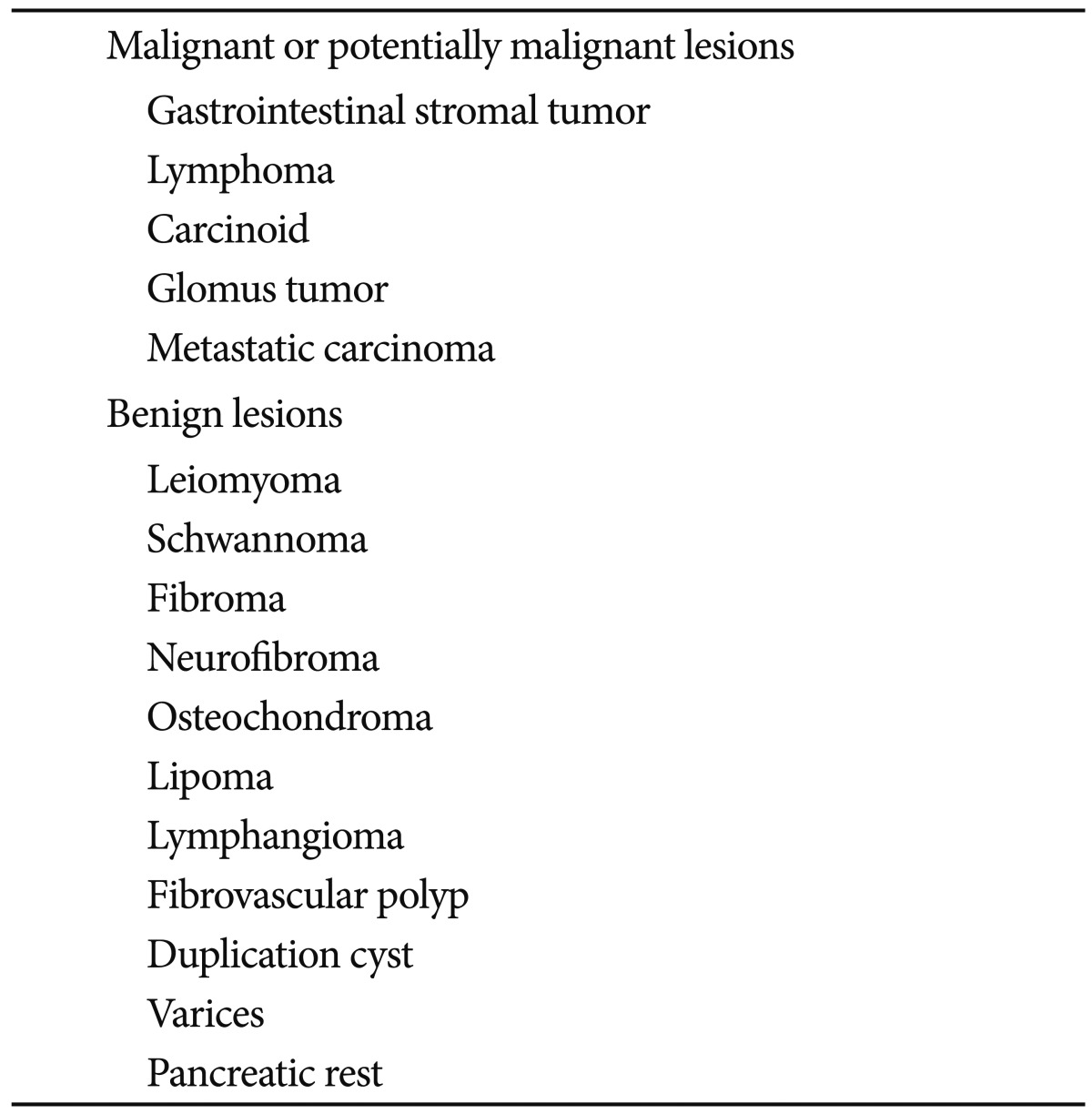
Subepithelial masses are a common endoscopic finding and are currently being encountered at an increasing rate given the widespread use of gastrointestinal (GI) endoscopy. The term subepithelial refers to a mass, bulge, or impression visible from the lumen arising from any layer of the GI wall or an adjacent structure causing extrinsic compression. The prevalence on routine endoscopy is unknown; however, one retrospective study reported subepithelial gastric lesions in 0.36% of esophagogastroduodenoscopies performed between 1976 and 1984.1 The differential diagnosis for these lesions is broad and includes benign, premalignant, malignant, and normal structures (Table 1). Treatment strategies vary accordingly, thus definitive diagnosis remains the utmost priority.
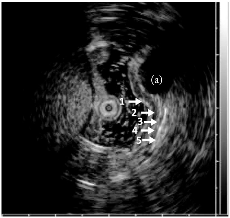
Endoscopic ultrasound (EUS) uses high frequency (5 to 20 MHz) sound waves to obtain detailed images of the GI tract wall and extraluminal structures and reliably differentiates between intramural wall lesions and extrinsic structures. EUS imaging of the GI tract typically identifies five distinct layers (Fig. 1), but can differentiate between seven or nine layers depending on the location of use and frequency of ultrasound probe. These layers have been histologically correlated.2 Characteristics of an intramural lesion including size, layer of origin, echotexture, and margins can be useful adjunctive information for accurate identification. Previous reports suggest extension between layers, irregular margins, or invasion into adjacent structures favor a malignant lesion aiding to guide treatment.3,4 However, endoscopic evaluation alone is insufficient for diagnosing the etiology of a subepithelial lesion.5 EUS has provided a major breakthrough for the characterization of subepithelial lesions, but histology is still needed in most situations.
Although rarely diagnostic, it is reasonable to perform biopsies of the mucosa overlying the subepithelial lesions.6 Stacked biopsies can be attempted; however, the yield remains low. Bite on bite technique using conventional sized forceps ranging from two to eight bites had a 38% diagnostic rate (54% in the esophagus, 28% in the stomach and duodenum) in a recent study.7 As such, techniques including endoscopic ultrasound-guided fine needle aspiration (EUS-FNA), EUS-guided Trucut biopsy (TCB), and EUS-guided fine needle biopsy (FNB) have been introduced to increase the diagnostic yield.
Pathologists have long preferred as much tissue as possible since the diagnosis typically requires immunohistochemical (IHC) staining for increased diagnostic yield. For subepithelial tumors (SETs), cytology, cell block processing, and IHC staining are all available using a 22-gauge EUS-FNA needle.8 IHC and mitotic counts usually cannot be performed on slides prepared for cytology; however, IHC can often be performed on cell blocks if sufficient quantities of cells have been collected.9 Therefore, a great deal of effort has been put forth to increase the diagnostic yield in subepithelial lesions, but each advancement continues to show its own distinct limitations.
EUS-FNA needles
EUS-FNA using 19-, 22-, or 25-gauge needles are commonly used to obtain tissue from suspicious lesions identified on EUS imaging. FNA with a 22-gauge needle can be used to obtain a cytological specimen with the occasional core tissue specimen by directing the needle into the lesion under ultrasound guidance (Fig. 2A).10 The sensitivity of EUS-FNA cytology for the diagnosis of GI stromal tumor was reported to be 78.4%; however, this sensitivity value was based on the identification of cells with spindle cell morphology and did not require confirmation with IHC.11 In this study, 62% of cases had a definitive diagnosis with IHC and 22% had a diagnosis that was suspicious for gastrointestinal stromal tumor with the finding of spindle cell morphology but not confirmed with IHC. IHC staining of various cell proteins can be performed on FNA samples if a sufficient quantity of cells is obtained to provide additional diagnostic information; however, critical architecture remains absent and a major drawback to FNA as a sole diagnostic procedure.
Recently, the Olympus Prototype Side-Port Needle (Olympus Corp., Tokyo, Japan) was developed in attempt to increase tissue acquisition and reduce the required number of overall passes for the diagnosis. In a pilot study, diagnostic material was obtained at the first pass in 56.2% of patients with the mean number of passes prior to diagnosis of 2.1. Overall, the diagnosis was reached in 94% of the patients. The safety profile was equivalent to similar products, but further prospective randomized trials are needed.12
Trucut core biopsy
To overcome the limitations of FNA, a 19-gauge Trucut core biopsy needle (QuickCore; Wilson-Cook Inc., Winston-Salem, NC, USA) has been proposed.13,14 This needle provides a core of tissue that can not only be used for individual cell morphology, but can be histologically examined for architectural change. Initial experience used in five patients with intramural lesions yielded the correct diagnosis in four of five cases compared to one of five cases using EUS-FNA.13 Some difficulties, such as needle stiffness, misfire of the needle inside the lesion and procedural difficulty when the lesion was in the distal antrum, were reported using EUS-TCB.15 A subsequent retrospective study looked at gastric SET greater than 2 cm in size. Nine were not able to be punctured and 19 more were unable to obtain adequate tissue. However, treatment plans were changed for 18 of 65 patients (27.7%) resulting in avoiding unnecessary resection, scheduling definitive treatment, and modifying the surgical field.16
Combination EUS-FNA with EUS-FNB
Both EUS-FNA and FNB have unique drawbacks limiting diagnostic accuracy. Combining these two techniques has been evaluated retrospectively with an overall accuracy of 95% (76% for EUS-FNA, 76% for EUS-FNB, 95% for combo; p=0.007) without an on site cytopathologist present.15 A prospective study followed demonstrating the yield of adequate tissue harvesting was similar for EUS-FNA and EUS-TCB (96.4% vs. 89.3%; p=not significant), with the same number of passes done. However, the accuracy for obtaining a specific diagnosis was significantly lower for EUS-FNA compared with EUS-TCB (5.3% and 68.4%; p<0.005). The information obtained was complementary, but EUS-TCB was useful when IHC was needed for definitive diagnosis of subepithelial mesenchymal tumors.17 A larger series followed showing EUS-FNA of subepithelial lesions was diagnostic in 61.6% and showed a spindle cell neoplasm (suspicious) in another 22.3% for an overall diagnostic yield of 83.9%. EUS-FNB did not increase the yield in this series.6 A randomized study comparing 22-gauge FNA and FNB needles in solid pancreatic masses further supported equal diagnostic sufficiency, technical performance, and safety profiles between the needles.18 In practice, combining EUS-FNA and EUS-FNB may not be practical due to the increased number of passes and higher cost. Instead, one method might be used as a rescue strategy when another one is failing.
ProCore needle
Due to limitations of the Trucut needle, a new histological needle with a core trap, a 19-gauge EUS-FNB device (ProCore; Cook Endoscopy, Winston-Salem, NC, USA), has been developed.19 This needle is uniquely designed to obtain both cytology and histology using reverse bevel technology, aiming for diagnosis on decreased number of passes. Core tissue samples can often be obtained using the ProCore needle making it possible to perform histology as well as IHC staining to help characterize SET (Fig. 2B). In a recent European study, the diagnostic accuracy was greater than 90% using this new 19-gauge EUS-FNB needle.20 The 22- and 25-gauge needles are also available.21 However, further studies are needed to validate this approach in subepithelial lesions.
EUS-FNA and core biopsy has emerged as a safe and effective diagnostic as well as therapeutic modality in recent years. The unique optical and mechanical properties require close attention during this procedure, but small series indicate the use of FNA needles by experienced operators does not carry an increased risk of complications.13,14,22,23 A learning curve exists for FNA, which may have a bearing on complications;24 however, the optimal number of procedure during training has not been defined. Theoretically, FNA or core biopsy of a subepithelial lesion carries a reduced complication rate when compared to transmural tissue acquisition.
For definitive diagnosis of subepithelial lesions, tissue acquisition from the lesion is necessary with both cytological and histochemical examinations including IHC stains. EUS-FNA is an effective method for obtaining tissue, but remains limited. EUS with Trucut appears to improve diagnostic accuracy, but is also limited by various factors including cumbersome use and variable yield. These two modalities should be considered complementary with the use of Trucut when IHC and tissue architectural details are required for diagnosis. Recently, EUS-guided core biopsy with a ProCore needle has become available. This allows core specimens with more tissue, IHC staining, and architecture, but more studies are needed to determine if the use of this particular needle improves the diagnostic yield in obtaining a tissue diagnosis in subepithelial lesions.
  • 1. Hedenbro JL, Ekelund M, Wetterberg P. Endoscopic diagnosis of submucosal gastric lesions. The results after routine endoscopy. Surg Endosc 1991;5:20–23.ArticlePubMed
  • 2. Kimmey MB, Martin RW, Haggitt RC, Wang KY, Franklin DW, Silverstein FE. Histologic correlates of gastrointestinal ultrasound images. Gastroenterology 1989;96(2 Pt 1):433–441.ArticlePubMed
  • 3. Chak A, Canto MI, Rösch T, et al. Endosonographic differentiation of benign and malignant stromal cell tumors. Gastrointest Endosc 1997;45:468–473.ArticlePubMed
  • 4. Palazzo L, Landi B, Cellier C, Cuillerier E, Roseau G, Barbier JP. Endosonographic features predictive of benign and malignant gastrointestinal stromal cell tumours. Gut 2000;46:88–92.ArticlePubMedPMC
  • 5. Hwang JH, Saunders MD, Rulyak SJ, Shaw S, Nietsch H, Kimmey MB. A prospective study comparing endoscopy and EUS in the evaluation of GI subepithelial masses. Gastrointest Endosc 2005;62:202–208.ArticlePubMed
  • 6. Hoda KM, Rodriguez SA, Faigel DO. EUS-guided sampling of suspected GI stromal tumors. Gastrointest Endosc 2009;69:1218–1223.ArticlePubMed
  • 7. Ji JS, Lee BI, Choi KY, et al. Diagnostic yield of tissue sampling using a bite-on-bite technique for incidental subepithelial lesions. Korean J Intern Med 2009;24:101–105.ArticlePubMedPMC
  • 8. Mekky MA, Yamao K, Sawaki A, et al. Diagnostic utility of EUS-guided FNA in patients with gastric submucosal tumors. Gastrointest Endosc 2010;71:913–919.ArticlePubMed
  • 9. Papanikolaou IS, Triantafyllou K, Kourikou A, Rösch T. Endoscopic ultrasonography for gastric submucosal lesions. World J Gastrointest Endosc 2011;3:86–94.ArticlePubMedPMC
  • 10. Matsui M, Goto H, Niwa Y, Arisawa T, Hirooka Y, Hayakawa T. Preliminary results of fine needle aspiration biopsy histology in upper gastrointestinal submucosal tumors. Endoscopy 1998;30:750–755.ArticlePubMedPDF
  • 11. Sepe PS, Moparty B, Pitman MB, Saltzman JR, Brugge WR. EUS-guided FNA for the diagnosis of GI stromal cell tumors: sensitivity and cytologic yield. Gastrointest Endosc 2009;70:254–261.ArticlePubMed
  • 12. Kaffes A, Corte C. Fine needle aspiration at endoscopic ultrasound with a novel side-port needle: a pilot experience. Therap Adv Gastroenterol 2012;5:89–94.ArticlePubMedPMC
  • 13. Levy MJ, Jondal ML, Clain J, Wiersema MJ. Preliminary experience with an EUS-guided trucut biopsy needle compared with EUS-guided FNA. Gastrointest Endosc 2003;57:101–106.ArticlePubMed
  • 14. Wiersema MJ, Levy MJ, Harewood GC, Vazquez-Sequeiros E, Jondal ML, Wiersema LM. Initial experience with EUS-guided trucut needle biopsies of perigastric organs. Gastrointest Endosc 2002;56:275–278.ArticlePubMed
  • 15. Storch I, Jorda M, Thurer R, et al. Advantage of EUS Trucut biopsy combined with fine-needle aspiration without immediate on-site cytopathologic examination. Gastrointest Endosc 2006;64:505–511.ArticlePubMed
  • 16. Lee JH, Choi KD, Kim MY, et al. Clinical impact of EUS-guided Trucut biopsy results on decision making for patients with gastric subepithelial tumors ≥ 2 cm in diameter. Gastrointest Endosc 2011;74:1010–1018.ArticlePubMed
  • 17. Saăftoiu A, Vilmann P, Guldhammer Skov B, Georgescu CV. Endoscopic ultrasound (EUS)-guided Trucut biopsy adds significant information to EUS-guided fine-needle aspiration in selected patients: a prospective study. Scand J Gastroenterol 2007;42:117–125.ArticlePubMed
  • 18. Bang JY, Hebert-Magee S, Trevino J, Ramesh J, Varadarajulu S. Randomized trial comparing the 22-gauge aspiration and 22-gauge biopsy needles for EUS-guided sampling of solid pancreatic mass lesions. Gastrointest Endosc 2012;76:321–327.ArticlePubMedPMC
  • 19. Polkowski M, Larghi A, Weynand B, et al. Learning, techniques, and complications of endoscopic ultrasound (EUS)-guided sampling in gastroenterology: European Society of Gastrointestinal Endoscopy (ESGE) technical guideline. Endoscopy 2012;44:190–206.ArticlePubMedPDF
  • 20. Iglesias-Garcia J, Poley JW, Larghi A, et al. Feasibility and yield of a new EUS histology needle: results from a multicenter, pooled, cohort study. Gastrointest Endosc 2011;73:1189–1196.ArticlePubMed
  • 21. Varadarajulu S, Fockens P, Hawes RH. Best practices in endoscopic ultrasound-guided fine-needle aspiration. Clin Gastroenterol Hepatol 2012;10:697–703.ArticlePubMed
  • 22. Larghi A, Verna EC, Stavropoulos SN, Rotterdam H, Lightdale CJ, Stevens PD. EUS-guided trucut needle biopsies in patients with solid pancreatic masses: a prospective study. Gastrointest Endosc 2004;59:185–190.ArticlePubMed
  • 23. Wildi SM, Hoda RS, Fickling W, et al. Diagnosis of benign cysts of the mediastinum: the role and risks of EUS and FNA. Gastrointest Endosc 2003;58:362–368.ArticlePubMed
  • 24. Eloubeidi MA, Tamhane A. EUS-guided FNA of solid pancreatic masses: a learning curve with 300 consecutive procedures. Gastrointest Endosc 2005;61:700–708.ArticlePubMed
Fig. 1
Endoscopic ultrasound imaging of the gastrointestinal tract wall. White arrows point to the corresponding histologic structures and echogenicity: 1) superficial mucosa (hyperechoic); 2) deep mucosa (hypoechoic); 3) submucosa (hyperechoic); 4) muscularis propria (hypoechoic); 5) serosa and subserosal fat (hyperechoic). (a) Subepithelial tumor continuous with the muscularis propria layer.
ce-46-441-g001.jpg
Fig. 2
Comparison of fine needle aspiration (FNA) and core biopsy samples. (A) FNA sample obtained with a 19-gauge needle. (B) Core biopsy sample obtained with a 19-gauge ProCore (Cook Endoscopy) needle (H&E stain, ×40).
ce-46-441-g002.jpg
Table 1
Differential Diagnosis of Gastric Subepithelial Lesions
ce-46-441-i001.jpg

Figure & Data

REFERENCES

    Citations

    Citations to this article as recorded by  
    • Endoscopic Management of Gastric Subepithelial Tumor
      Hyunchul Lim
      Journal of Digestive Cancer Research.2022; 10(1): 16.     CrossRef
    • Magnetically Actuated Soft Capsule Endoscope for Fine-Needle Biopsy
      Donghoon Son, Hunter Gilbert, Metin Sitti
      Soft Robotics.2020; 7(1): 10.     CrossRef
    • Endoscopic ultrasound guided fine‐needle aspiration vs core needle biopsy for solid pancreatic lesions: Comparison of diagnostic accuracy and procedural efficiency
      Aslam Syed, Olivia Babich, Bharat Rao, Shailendra Singh, Neil Carleton, Abhishek Gulati, Archana Kulkarni, Mrinal Garg, Katie Farah, Gursimran Kochhar, Suzanne Morrissey, Marcia Mitre, Abhijit Kulkarni, Manish Dhawan, Jan F. Silverman, Majed Pharaon, Shya
      Diagnostic Cytopathology.2019; 47(11): 1138.     CrossRef
    • Cytology and histology: Complementary diagnostic modalities during endoscopic ultrasound-guided tissue acquisition
      Norge Vergara, Roseann I. Wu, Stuti Shroff, Cindy M. McGrath
      Techniques in Gastrointestinal Endoscopy.2018; 20(1): 10.     CrossRef
    • Echoendoscopic ablative therapy for solid pancreatic tumors
      Woo Hyun Paik, Dong Wan Seo
      Journal of Digestive Diseases.2017; 18(3): 135.     CrossRef
    • Via mucosa incision EUS-guided sampling for the diagnosis of conventional endoscopic biopsy-negative gastric wall thickening
      Hongbo Shan, Xiaoyan Gao, Guangyu Luo, Jieqing Xiang, Bilv Zhong, Xiaofang Qiu, Shiyong Lin, Shuhong Li, Yin Li, Guoliang Xu, Rong Zhang
      Scientific Reports.2017;[Epub]     CrossRef
    • Diagnosis of Gastric Subepithelial Tumor: Focusing on Endoscopic Ultrasonography
      Eun Young Kim
      The Korean Journal of Helicobacter and Upper Gastrointestinal Research.2015; 15(1): 9.     CrossRef
    • Which Needle Is Better for Diagnosing Subepithelial Lesions?
      Eun Young Kim
      Clinical Endoscopy.2015; 48(2): 91.     CrossRef

    • PubReader PubReader
    • ePub LinkePub Link
    • Cite
      CITE
      export Copy Download
      Close
      Download Citation
      Download a citation file in RIS format that can be imported by all major citation management software, including EndNote, ProCite, RefWorks, and Reference Manager.

      Format:
      • RIS — For EndNote, ProCite, RefWorks, and most other reference management software
      • BibTeX — For JabRef, BibDesk, and other BibTeX-specific software
      Include:
      • Citation for the content below
      Endoscopic Ultrasound-Fine Needle Aspiration versus Core Biopsy for the Diagnosis of Subepithelial Tumors
      Clin Endosc. 2013;46(5):441-444.   Published online September 30, 2013
      Close
    • XML DownloadXML Download
    Figure
    • 0
    • 1
    Endoscopic Ultrasound-Fine Needle Aspiration versus Core Biopsy for the Diagnosis of Subepithelial Tumors
    Image Image
    Fig. 1 Endoscopic ultrasound imaging of the gastrointestinal tract wall. White arrows point to the corresponding histologic structures and echogenicity: 1) superficial mucosa (hyperechoic); 2) deep mucosa (hypoechoic); 3) submucosa (hyperechoic); 4) muscularis propria (hypoechoic); 5) serosa and subserosal fat (hyperechoic). (a) Subepithelial tumor continuous with the muscularis propria layer.
    Fig. 2 Comparison of fine needle aspiration (FNA) and core biopsy samples. (A) FNA sample obtained with a 19-gauge needle. (B) Core biopsy sample obtained with a 19-gauge ProCore (Cook Endoscopy) needle (H&E stain, ×40).
    Endoscopic Ultrasound-Fine Needle Aspiration versus Core Biopsy for the Diagnosis of Subepithelial Tumors

    Differential Diagnosis of Gastric Subepithelial Lesions

    Table 1 Differential Diagnosis of Gastric Subepithelial Lesions


    Clin Endosc : Clinical Endoscopy Twitter Facebook
    Close layer
    TOP