Skip Navigation
Skip to contents

Clin Endosc : Clinical Endoscopy

OPEN ACCESS

Articles

Page Path
HOME > Clin Endosc > Volume 52(2); 2019 > Article
Focused Review Series: Endoscopic Submucosal Dissection for Colorectal Tumors Estimation of Invasion Depth: The First Key to Successful Colorectal ESD
Bo-In Lee1,2, Takahisa Matsuda3,orcid
Clinical Endoscopy 2019;52(2):100-106.
DOI: https://doi.org/10.5946/ce.2019.012
Published online: March 27, 2019

1Division of Gastroenterology, Department of Internal Medicine, College of Medicine, The Catholic University of Korea, Seoul, Korea

2Division of Gastroenterology, Department of Internal Medicine, College of Medicine, Catholic Photomedicine Research Institute, Seoul, Korea

3Cancer Screening Center/Endoscopy Division, National Cancer Center Hospital, Division of Screening Technology, Center for Public Health Sciences, National Cancer Center, Tokyo, Japan

Correspondence: Takahisa Matsuda Cancer Screening Center/Endoscopy Division, National Cancer Center Hospital, Division of Screening Technology, Center for Public Health Sciences, National Cancer Center, 5-1-1 Tsukiji, Chuo-ku, Tokyo 104-0045, Japan Tel: +81-3-3542-2511, Fax: +81-3-3547-6125, E-mail: tamatsud@ncc.go.jp
• Received: December 10, 2018   • Revised: March 4, 2019   • Accepted: March 4, 2019

Copyright © 2019 Korean Society of Gastrointestinal Endoscopy

This is an Open Access article distributed under the terms of the Creative Commons Attribution Non-Commercial License (http://creativecommons.org/licenses/by-nc/3.0/) which permits unrestricted non-commercial use, distribution, and reproduction in any medium, provided the original work is properly cited.

  • 8,416 Views
  • 358 Download
  • 20 Web of Science
  • 19 Crossref
  • 19 Scopus
prev next
  • Colorectal tumors with superficial submucosal invasion, which cannot be removed by snaring, are one of the most optimal indications for colorectal endoscopic submucosal dissection (ESD). Therefore, estimation of the invasion depth is the first key to successful colorectal ESD. Although estimation of the invasion depth based on the gross morphology may be useful in selected cases, its diagnostic accuracy could not reach the clinical requirement. The Japan Narrow-band Imaging (NBI) Expert Team (JNET) classification of NBI magnifying endoscopy findings is a useful method for histologic prediction and invasion depth estimation. However, magnifying chromoendoscopy is still necessary for JNET type 2B lesions to reach a satisfactory diagnostic accuracy. Endocytoscopy with artificial intelligence is a promising technology in invasion depth estimation; however, more data are needed for its clinical application.
Intramucosal colorectal cancers without lymph node metastasis are good indications for endoscopic treatment [1-3]. Lymph node metastasis can be truly determined only by histopathologic examination of the surgical specimen, whereas endoscopic assessment allows only estimating the risk of invasion and metastasis [4]. Unfavorable histologic findings including submucosal invasion 1,000 μm, positive lymphovascular invasion, poorly differentiated adenocarcinoma, signet-ring cell carcinoma, mucinous carcinoma, or grade 2/3 budding at the site of the deepest invasion are considered risk factors of lymph node metastasis, requiring radical dissection or systemic treatment [5,6]. Therefore, estimation of the invasion depth is one of the most important steps in establishing the treatment strategies for colorectal cancer.
Colorectal endoscopic submucosal dissection (ESD) can achieve en bloc resection in >80% of the indicated cases [7]. However, it is still a technically challenging, time-consuming, and resource-consuming procedure, in addition to its high risk of adverse events.
The current indications for colorectal ESD vary among different countries. The Korean Heath Insurance Review and Assessment Service announced that the costs of colorectal ESD can be reimbursed for a well-differentiated mucosal cancer ≤5 cm without ulceration, a laterally spreading tumor (LST) ≥2 cm, a sessile polyp ≥2 cm, and an adenoma with fibrosis. The Japanese Colorectal ESD Standardization Implementation Working Group proposed that colorectal ESD should be indicated in lesions for which en bloc resection with snare endoscopic mucosal resection (EMR) is difficult to apply (LSTnongranular [LST-NG] particularly the LST-NG, pseudodepressed type, lesions showing a VI‐type pit pattern, carcinoma with shallow T1 [submucosa] invasion, large depressed‐type tumors, and large protruded‐type lesions suspected to be carcinoma), mucosal tumors with submucosal fibrosis, sporadic localized tumors in chronic inflammatory conditions such as ulcerative colitis, and local residual or recurrent early carcinomas after endoscopic resection [8,9]. The European Society of Gastrointestinal Endoscopy also proposed that ESD can be considered for the removal of colonic and rectal lesions with a high suspicion of limited submucosal invasion, based on the 2 main criteria of depressed morphology and irregular or nongranular surface pattern (particularly if the lesions are >20 mm), or that ESD can be considered for colorectal lesions that otherwise cannot be optimally and radically removed using snare-based techniques [10]. All of the indications include tumors with superficial submucosal invasion as good candidates for colorectal ESD. Recently, an Australian group suggested that performing ESD only for lesions highly suspicious for containing submucosal invasive cancer is the most cost-effective strategy for removing large sessile tumors and LSTs >2 cm [11]. All the indications and studies suggest that estimation of the depth of invasion should be the priority when considering whether a colorectal neoplasm could be treated with endoscopic resection including ESD.
The depth of invasion can be estimated using the gross findings of white-light endoscopy. Size is one of the most important gross findings suggesting submucosal invasion. Submucosal invasion was found in 7.4%–14% of colorectal polyps >2 cm in a Japanese study [12]. Other endoscopic features related to deep submucosal invasion were reported as follows: loss of lobulation, excavation, demarcated depressed area, stalk swelling, fullness, and fold convergence [13,14]. However, multivariate analysis revealed only loss of lobulation in the sessile type and fullness in the superficial type as the independent risk factors of deep submucosal invasion [13]. According to a Korean study including 39 superficial submucosal cancers and 25 deep submucosal cancers <2 cm, the positive predictive value and diagnostic accuracy for deep submucosal invasion of gross morphology assessment were 0.27 and 0.71, respectively, which are insufficient for clinical application [14]. The diagnostic accuracy of the 3 Korean experts who investigated the gross morphology were 74%–82% for 15 submucosal cancers and 35 mucosal cancers, and their agreement was not high (κ=0.298–0.649) [15].
Although estimation of the invasion depth based on gross features may be simple, less resource consuming, and useful in selected cases, its diagnostic accuracy and interobserver agreement still seems insufficient for clinical application.
Until 2009, several Japanese academic centers proposed their narrow-band Imaging (NBI) classifications (Sano, Hiroshima, Showa, and Jikei) based on the microvasculature pattern [16-19]. These classifications were used for histologic prediction and invasion depth estimation. Even a brief education on NBI magnifying endoscopy findings can improve trainees’ accuracy in estimating the invasion depth [20]. However, there were concerns about the complicated classifications. In 2009, the Colon Tumor NBI Interest Group, which consisted of 6 experts from Japan, the United States, France, and the United Kingdom, proposed the NBI International Colorectal Endoscopic (NICE) classification. This classification is based on the color and the vascular and surface pattern of tumors, which can be assessed using nonmagnifying endoscopy. International studies have proved that this simple system is also useful for predicting the histology and estimating the invasion depth of colorectal lesions [15,21,22].
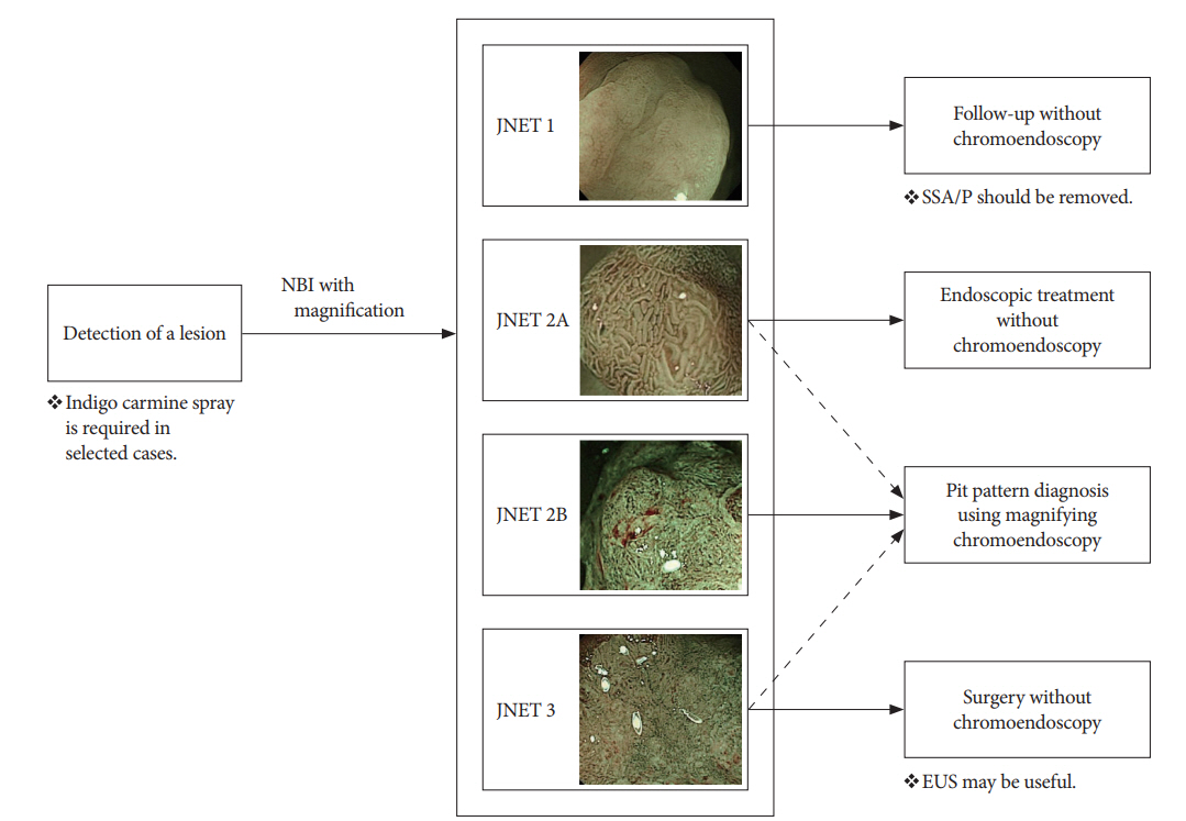
However, NBI findings suggesting superficial submucosal invasive cancer were not described in the NICE classification. In 2014, the Japan NBI Expert Team (JNET), comprising 38 Japanese specialists, proposed the JNET classification to unify the previous classifications of NBI magnifying endoscopy findings [23]. This classification was established to provide not only histologic prediction but also estimation of the invasion depth with NBI magnifying endoscopy [24,25]. This classification has 4 categories according to the vascular and surface pattern: JNET type 1, type 2A, type 2B, and type 3. Type 1 usually suggests hyperplastic polyp (HP) or sessile serrated polyp (SSP); type 2A is compatible with low-grade intramucosal neoplasms; type 2B includes high-grade intramucosal neoplasia or shallow submucosal invasive cancer; and type 3 means deep submucosal invasive cancer (Table 1) [23].
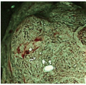
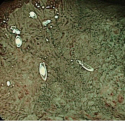
According to the Japanese studies that followed the JNET proposal, the classification was useful for the diagnosis of HP/SSP (type 1), low-grade dysplasia (type 2A), and deep submucosal cancer (type 3), but not for high-grade dysplasia/superficial submucosal cancer (type 2B). A retrospective image evaluation study revealed that the sensitivity, specificity, positive predictive value, negative predictive value, and diagnostic accuracy for type 2B lesions were 75.6%, 90.5%, 67.3%, 93.4%, and 87.4%, respectively [26]. Another study also showed that the specificity and positive predictive value of type 2B lesions judged by 3 expert endoscopists were 82.8% and 50.9%, which were lower than those of other type lesions [27]. Of lesions diagnosed as type 2B, 37% were low-grade adenomas and 12% were deep invasive cancers. Therefore, the authors proposed an additional evaluation, such as pit pattern diagnosis using chromoendoscopy, for the accurate diagnosis of type 2B lesions (Fig. 1) [27]. More data are required to evaluate how much the diagnostic accuracy for JNET type 2B lesions can be improved by additional magnifying chromoendoscopy.
The surface of the lesion is washed with proteinase to remove overlying mucous for pit pattern diagnosis, after which indigo carmine solution is sprayed. When magnifying endoscopy with indigo carmine is not enough for an accurate pit pattern diagnosis, crystal violet solution is applied as a staining agent. However, the application of crystal violet may be limited outside of Japan because of the potential genotoxicity of this agent [28].
Studies on pit pattern diagnosis with magnifying chromoendoscopy have confirmed that specific categories of pit patterns are suggestive of submucosal invasive cancer. A study conducted at the Northern Yokohama Hospital showed that 34% of the lesions with a category VI (irregular) pit pattern and 91% of the lesions with a category VN (nonstructural/amorphous) pattern were submucosal cancers, whereas only 0.64% of pit pattern III or IV were submucosal cancers [12]. As mentioned above, the type VI pattern is associated with a variety of lesions, such as low-grade dysplasia, high-grade dysplasia/superficial submucosal cancer, and even deep submucosal cancer [29,30].
Therefore, there have been trials classifying the type VI pattern into subtypes for better prediction of invasion depth. Onishi et al. suggested that the following 4 patterns are suggestive of deep submucosal invasion: absence of pits that can be recognized as having originated from type III or IV, marginal irregularities of the gland duct, narrowing of the gland duct lumen, and unclear outline of the gland duct [29]. Matsuda et al. reported that the sensitivity, specificity, positive predictive value, negative predictive value, and diagnostic accuracy for 3,371 adenomas, 612 intramucosal cancers, 52 superficial submucosal cancers and 180 deep submucosal cancers were 85.6%, 99.4%, 86.5%, 99.4%, and 98.8%, respectively, when VI lesions with a demarcated area (clearly visualized zone between 2 morphologically different types of pits, e.g., depression, large nodule, or reddened area) were considered to have a deep submucosal invasive pattern [3]. Kanao et al. subclassified VI lesions into those with mild irregularity and those with severe irregularity (unclear staining between pits, irregular pit margins, and VI area ≥5 mm) [30]. They found that 56.1% of severely irregular lesions were deep submucosal invasive cancers, whereas 6.7% of mildly irregular lesions were deep submucosal invasive cancers [30].
As insertion of an echoendoscope into the colonic lumen is not feasible, a high-frequency device (20-MHz miniprobe) is generally used to determine the depth of invasion. A hypoechoic area breaking into the third layer is usually considered to indicate deep submucosal invasion. As the penetration depth of a high-frequency probe is limited, it cannot be used for the evaluation of the invasion depth of a large sessile or pedunculated lesion.
The current data on the diagnostic accuracy of endoscopic ultrasonography (EUS) for estimating the invasion depth are conflicting. Two previous trials that were performed prospectively suggested that EUS was superior to magnifying chromoendoscopy, with a higher accuracy of invasion depth determination (92% vs. 63% [31] and 93% vs. 59% [32]). However, both studies had a limitation: when performing EUS, the endoscopists had more information because EUS was performed after magnifying chromoendoscopy. A recent prospective trial, with a cross-over design, on magnifying chromoendoscopy and EUS revealed that the diagnostic accuracy for depth of invasion was comparable between the two methods (71% vs. 71%); however, EUS required a significantly longer procedure time than magnifying chromoendoscopy, and the authors concluded that neither procedure has sufficient diagnostic accuracy [33].
Ultra-high magnification images (×450) can be obtained using an endocytoscope after staining with 0.05% crystal violet and 1.0 methylene blue, which stains cellular matrices and nuclei, respectively. In a pilot study, the diagnostic accuracy of endocytoscopy for deep invasive submucosal cancer was 95.9% [34]. Endocytoscopy images were also interpreted using artificial intelligence, and the algorithm obtained after 5,543 images from 238 lesions could diagnose invasive colorectal cancer in 200 test images with an accuracy of 94.1% [35].
Although noninvasive neoplasia can be resected using EMR or piecemeal EMR regardless of size, ESD can also be attempted for noninvasive lesions ≥2 cm, especially in experienced institutions, to accomplish en bloc resection and to decrease local recurrence. When a lesion with suspected superficial submucosal invasion cannot be optimally and radically removed using a snare-based technique, en bloc resection with ESD is strongly recommended. Surgical resection is definitely recommended for lesions for which there is high confidence about the presence of deep submucosal invasion [36].
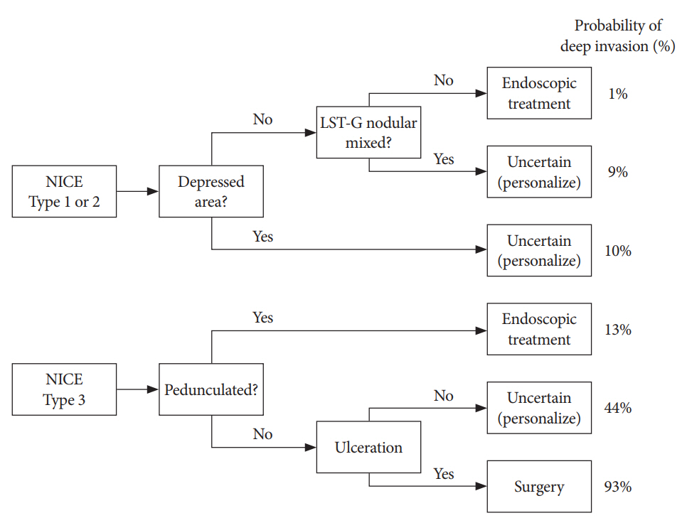
In a Spanish study analyzing 2,123 colon lesions >1 cm, a pedunculated morphology, ulceration, depressed area, or nodular mixed morphology affected the diagnostic accuracy of the NICE classification. Moreover, there remained subgroups of lesions for which endoscopic estimation of the invasion depth was difficult, including NICE type 2 with depressed area, NICE type 2 nodular mixed type, and NICE type 3 nonpedunculated polyps without ulceration. The authors proposed a management algorithm for polyps >1 cm according to the probability of deep invasion and endoscopic resectability (Fig. 2) [37].
Even with all the currently available modalities for estimating the invasion depth before colorectal ESD, including the assessment of gross features, NBI, and magnifying chromoendoscopy, a perfect estimation is not possible; thus, the clinical decision after colorectal ESD should be mainly based on the final histopathologic results. As mentioned earlier, additional surgery should be considered when the resected specimen shows unfavorable histologic findings [5,6].
Estimation of the invasion depth is an important step for successful colorectal ESD. Currently, NBI magnifying endoscopy is one of the most useful and practical methods of estimating the invasion depth of colorectal tumors. Although JNET type 2B (irregular vessel and surface pattern) represents high-grade intramucosal neoplasia/shallow submucosal invasive cancer, it also occasionally suggests deep submucosal invasive cancer or low-grade intramucosal neoplasm. Magnifying chromoendoscopy is recommended for JNET type 2B lesions, and more studies are required to elucidate how much diagnostic accuracy can be improved by additional magnifying chromoendoscopy.
Fig. 1.
Strategy for the endoscopic treatment of colorectal lesions according to the Japan Narrow-band Imaging (NBI) Expert Team (JNET) classification. The dotted arrows suggest that magnifying chromoendoscopy for pit pattern diagnosis can be performed in selected cases of JNET 2A or 3 with low confidence. EUS, endoscopic ultrasonography; SSA/P, sessile serrated adenoma/polyp (Adapted and modified from Sumimoto et al. [37].
ce-2019-012f1.jpg
Fig. 2.
Proposed treatment algorithm based on the probability of deep invasion and endoscopic resectability (Adapted and modified from Puig et al. [37]. LST-G, laterally spreading tumor-granular; NICE, narrow-band imaging international colorectal endoscopic.
ce-2019-012f2.jpg
Table 1.
The Japan Narrow-Band Imaging Expert Team Classification (Adapted and modified from Sano et al. [23]
Type 1 Type 2A Type 2B Type 3
NICE classification Type 1 Type 2 Type 3
Vessel pattern Invisiblea) Regular caliber Variable caliber Loose vessel areas
Regular distribution (meshed/spiral pattern)b) Irregular distribution Interruption of thick vessels
Surface pattern Regular dark or white spots Regular (tubular/branched/papillary) Irregular or obscure Amorphous areas
Similar to surrounding normal mucosa
Most likely histology Hyperplastic polyp/ Sessile serrated polyp Low grade intramucosal neoplasm High grade intramucosal neoplasm/ Shallow submucosal invasive cancerc) Deep submucosal invasive cancer
Endoscopic images ce-2019-012i1.jpg ce-2019-012i2.jpg ce-2019-012i3.jpg ce-2019-012i4.jpg

NICE, narrow-band imaging international colorectal endoscopic.

a)If visible, the caliber in the lesion is similar to surrounding normal mucosa.

b)Microvessels are often distributed in a punctate pattern and well-ordered reticular or spiral vessels may not be observed in depressed lesions.

c)Deep submucosal invasive cancer may be included.

  • 1. Morson BC, Whiteway JE, Jones EA, Macrae FA, Williams CB. Histopathology and prognosis of malignant colorectal polyps treated by endoscopic polypectomy. Gut 1984;25:437–444.ArticlePubMedPMC
  • 2. Fujimori T, Kawamata H, Kashida H. Precancerous lesions of the colorectum. J Gastroenterol 2001;36:587–594.ArticlePubMedPDF
  • 3. Matsuda T, Fujii T, Saito Y, et al. Efficacy of the invasive/non-invasive pattern by magnifying chromoendoscopy to estimate the depth of invasion of early colorectal neoplasms. Am J Gastroenterol 2008;103:2700–2706.ArticlePubMed
  • 4. Hewett DG, Sakata S. Classifications for optical diagnosis of colorectal lesions: not 2B with JNET. Gastrointest Endosc 2017;85:822–828.ArticlePubMed
  • 5. Watanabe T, Itabashi M, Shimada Y, et al. Japanese Society for Cancer of the Colon and Rectum (JSCCR) guidelines 2014 for treatment of colorectal cancer. Int J Clin Oncol 2015;20:207–239.ArticlePubMedPMC
  • 6. Kitajima K, Fujimori T, Fujii S, et al. Correlations between lymph node metastasis and depth of submucosal invasion in submucosal invasive colorectal carcinoma: a Japanese collaborative study. J Gastroenterol 2004;39:534–543.ArticlePubMedPDF
  • 7. Repici A, Hassan C, De Paula Pessoa D, et al. Efficacy and safety of endoscopic submucosal dissection for colorectal neoplasia: a systematic review. Endoscopy 2012;44:137–150.ArticlePubMedPDF
  • 8. Tanaka S, Kashida H, Saito Y, et al. JGES guidelines for colorectal endoscopic submucosal dissection/endoscopic mucosal resection. Dig Endosc 2015;27:417–434.ArticlePubMed
  • 9. Tanaka S, Oka S, Chayama K. Colorectal endoscopic submucosal dissection: present status and future perspective, including its differentiation from endoscopic mucosal resection. J Gastroenterol 2008;43:641–651.ArticlePubMedPDF
  • 10. Pimentel-Nunes P, Dinis-Ribeiro M, Ponchon T, et al. Endoscopic submucosal dissection: European Society of Gastrointestinal Endoscopy (ESGE) guideline. Endoscopy 2015;47:829–854.ArticlePubMedPDF
  • 11. Bahin FF, Heitman SJ, Rasouli KN, et al. Wide-field endoscopic mucosal resection versus endoscopic submucosal dissection for laterally spreading colorectal lesions: a cost-effectiveness analysis. Gut 2018;67:1965–1973.ArticlePubMed
  • 12. Kudo S, Lambert R, Allen JI, et al. Nonpolypoid neoplastic lesions of the colorectal mucosa. Gastrointest Endosc 2008;68(4 Suppl):S3–S47.ArticlePubMed
  • 13. Ikehara H, Saito Y, Matsuda T, Uraoka T, Murakami Y. Diagnosis of depth of invasion for early colorectal cancer using magnifying colonoscopy. J Gastroenterol Hepatol 2010;25:905–912.ArticlePubMed
  • 14. Park W, Kim B, Park SJ, et al. Conventional endoscopic features are not sufficient to differentiate small, early colorectal cancer. World J Gastroenterol 2014;20:6586–6593.ArticlePubMedPMC
  • 15. Choi HJ, Lee BI, Choi H, et al. Diagnostic accuracy and interobserver agreement in predicting the submucosal invasion of colorectal tumors using gross findings, pit patterns, and microvasculatures. Clin Endosc 2013;46:168–171.ArticlePubMedPMCPDF
  • 16. Sano Y, Ikematsu H, Fu KI, et al. Meshed capillary vessels by use of narrow-band imaging for differential diagnosis of small colorectal polyps. Gastrointest Endosc 2009;69:278–283.ArticlePubMed
  • 17. Kanao H, Tanaka S, Oka S, Hirata M, Yoshida S, Chayama K. Narrow-band imaging magnification predicts the histology and invasion depth of colorectal tumors. Gastrointest Endosc 2009;69(3 Pt 2):631–636.ArticlePubMed
  • 18. Wada Y, Kudo SE, Kashida H, et al. Diagnosis of colorectal lesions with the magnifying narrow-band imaging system. Gastrointest Endosc 2009;70:522–531.ArticlePubMed
  • 19. Aihara H, Saito S, Tajiri H. Rationale for and clinical benefits of colonoscopy with narrow band imaging: pathological prediction and colorectal screening. Int J Colorectal Dis 2013;28:1–7.ArticlePubMedPDF
  • 20. Kim JS, Lee BI, Choi H, et al. Brief education on microvasculature and pit pattern for trainees significantly improves estimation of the invasion depth of colorectal tumors. Gastroenterol Res Pract 2014;2014:245396.ArticlePubMedPMCPDF
  • 21. Hewett DG, Kaltenbach T, Sano Y, et al. Validation of a simple classification system for endoscopic diagnosis of small colorectal polyps using narrow-band imaging. Gastroenterology 2012;143:599–607.e1.ArticlePubMed
  • 22. Hayashi N, Tanaka S, Hewett DG, et al. Endoscopic prediction of deep submucosal invasive carcinoma: validation of the narrow-band imaging international colorectal endoscopic (NICE) classification. Gastrointest Endosc 2013;78:625–632.ArticlePubMed
  • 23. Sano Y, Tanaka S, Kudo SE, et al. Narrow-band imaging (NBI) magnifying endoscopic classification of colorectal tumors proposed by the Japan NBI Expert Team. Dig Endosc 2016;28:526–533.ArticlePubMed
  • 24. Sano Y, Hirata D, Saito Y. Japan NBI Expert Team classification: narrow-band imaging magnifying endoscopic classification of colorectal tumors. Dig Endosc 2018;30:543–545.ArticlePubMed
  • 25. Iwatate M, Sano Y, Tanaka S, et al. Validation study for development of the Japan NBI Expert Team classification of colorectal lesions. Dig Endosc 2018;30:642–651.ArticlePubMed
  • 26. Komeda Y, Kashida H, Sakurai T, et al. Magnifying narrow band imaging (NBI) for the diagnosis of localized colorectal lesions using the Japan NBI Expert Team (JNET) classification. Oncology 2017;93 Suppl 1:49–54.ArticlePubMed
  • 27. Sumimoto K, Tanaka S, Shigita K, et al. Clinical impact and characteristics of the narrow-band imaging magnifying endoscopic classification of colorectal tumors proposed by the Japan NBI Expert Team. Gastrointest Endosc 2017;85:816–821.ArticlePubMed
  • 28. Mani S, Bharagava RN. Exposure to crystal violet, its toxic, genotoxic and carcinogenic effects on environment and its degradation and detoxification for environmental safety. Rev Environ Contam Toxicol 2016;237:71–104.ArticlePubMed
  • 29. Onishi T, Tamura S, Kuratani Y, Onishi S, Yasuda N. Evaluation of the depth score of type V pit patterns in crypt orifices of colorectal neoplastic lesions. J Gastroenterol 2008;43:291–297.ArticlePubMedPDF
  • 30. Kanao H, Tanaka S, Oka S, et al. Clinical significance of type V(I) pit pattern subclassification in determining the depth of invasion of colorectal neoplasms. World J Gastroenterol 2008;14:211–217.ArticlePubMedPMC
  • 31. Matsumoto T, Hizawa K, Esaki M, et al. Comparison of EUS and magnifying colonoscopy for assessment of small colorectal cancers. Gastrointest Endosc 2002;56:354–360.ArticlePubMed
  • 32. Hurlstone DP, Brown S, Cross SS, Shorthouse AJ, Sanders DS. High magnification chromoscopic colonoscopy or high frequency 20 MHz mini probe endoscopic ultrasound staging for early colorectal neoplasia: a comparative prospective analysis. Gut 2005;54:1585–1589.ArticlePubMedPMC
  • 33. Shimura T, Ebi M, Yamada T, et al. Magnifying chromoendoscopy and endoscopic ultrasonography measure invasion depth of early stage colorectal cancer with equal accuracy on the basis of a prospective trial. Clin Gastroenterol Hepatol 2014;12:662–668; e1-e2.ArticlePubMed
  • 34. Kudo SE, Wakamura K, Ikehara N, Mori Y, Inoue H, Hamatani S. Diagnosis of colorectal lesions with a novel endocytoscopic classification - a pilot study. Endoscopy 2011;43:869–875.ArticlePubMedPDF
  • 35. Takeda K, Kudo SE, Mori Y, et al. Accuracy of diagnosing invasive colorectal cancer using computer-aided endocytoscopy. Endoscopy 2017;49:798–802.ArticlePubMedPDF
  • 36. Nishizawa T, Yahagi N. Endoscopic mucosal resection and endoscopic submucosal dissection: technique and new directions. Curr Opin Gastroenterol 2017;33:315–319.ArticlePubMed
  • 37. Puig I, López-Cerón M, Arnau A, et al. Accuracy of the narrow-band imaging international colorectal endoscopic classification system in identification of deep invasion in colorectal polyps. Gastroenterology 2019;156:75–87.ArticlePubMed

Figure & Data

REFERENCES

    Citations

    Citations to this article as recorded by  
    • Deep Learning in Endoscopic Ultrasound: A Breakthrough in Detecting Distal Cholangiocarcinoma
      Rares Ilie Orzan, Delia Santa, Noemi Lorenzovici, Thomas Andrei Zareczky, Cristina Pojoga, Renata Agoston, Eva-Henrietta Dulf, Andrada Seicean
      Cancers.2024; 16(22): 3792.     CrossRef
    • Comparison of two pathological processing methods for large endoscopic submucosal dissection (ESD) specimens
      Zixiang Yu, Dongxian Jiang, Wen Huang, Rongkui Luo, Haixing Wang, Jieakesu Su, Jia Liu, Chen Xu, Yingyong Hou
      Journal of Clinical Pathology.2023; 76(11): 757.     CrossRef
    • Endoscopic submucosal dissection for colorectal polyps: outcome determining factors
      Chi Woo Samuel Chow, Tak Lit Derek Fung, Pak Tat Chan, Kam Hung Kwok
      Surgical Endoscopy.2023; 37(2): 1293.     CrossRef
    • Endoscopic management of patients with high-risk colorectal colitis–associated neoplasia: a Delphi study
      Michiel T.J. Bak, Eduardo Albéniz, James E. East, Nayantara Coelho-Prabhu, Noriko Suzuki, Yutaka Saito, Takayuki Matsumoto, Rupa Banerjee, Michal F. Kaminski, Ralf Kiesslich, Emmanuel Coron, Annemarie C. de Vries, C. Janneke van der Woude, Raf Bisschops,
      Gastrointestinal Endoscopy.2023; 97(4): 767.     CrossRef
    • Detecting colorectal lesions with image-enhanced endoscopy: an updated review from clinical trials
      Mizuki Nagai, Sho Suzuki, Yohei Minato, Fumiaki Ishibashi, Kentaro Mochida, Ken Ohata, Tetsuo Morishita
      Clinical Endoscopy.2023; 56(5): 553.     CrossRef
    • Development of artificial intelligence technology in diagnosis, treatment, and prognosis of colorectal cancer
      Feng Liang, Shu Wang, Kai Zhang, Tong-Jun Liu, Jian-Nan Li
      World Journal of Gastrointestinal Oncology.2022; 14(1): 124.     CrossRef
    • Linear-array EUS improves the accuracy of predicting deep submucosal invasion in non-pedunculated rectal polyps compared with radial EUS: a prospective observational study
      Zhixian Lan, Kangyue Sun, Yuchen Luo, Haiyan Hu, Wei Zhu, Wen Guo, Jing Wen, Wenting Mi, Junsheng Chen, Xiang Chen, Venkata Akshintala, Ying Huang, Side Liu, Yue Li
      Surgical Endoscopy.2021; 35(4): 1734.     CrossRef
    • Deep learning analysis for the detection of pancreatic cancer on endosonographic images: a pilot study
      Ryosuke Tonozuka, Takao Itoi, Naoyoshi Nagata, Hiroyuki Kojima, Atsushi Sofuni, Takayoshi Tsuchiya, Kentaro Ishii, Reina Tanaka, Yuichi Nagakawa, Shuntaro Mukai
      Journal of Hepato-Biliary-Pancreatic Sciences.2021; 28(1): 95.     CrossRef
    • Comparison of long-term recurrence-free survival between primary surgery and endoscopic resection followed by secondary surgery in T1 colorectal cancer
      Eun Hye Oh, Nayoung Kim, Sung Wook Hwang, Sang Hyoung Park, Dong-Hoon Yang, Byong Duk Ye, Seung-Jae Myung, Suk-Kyun Yang, Chang Sik Yu, Jin Cheon Kim, Jeong-Sik Byeon
      Gastrointestinal Endoscopy.2021; 94(2): 394.     CrossRef
    • The impact of transanal local excision of early rectal cancer on completion rectal resection without neoadjuvant chemoradiotherapy: a systematic review
      R. Zinicola, R. Nascimbeni, R. Cirocchi, G. Gagliardi, N. Cracco, M. Giuffrida, G. Pedrazzi, G. A. Binda
      Techniques in Coloproctology.2021; 25(9): 997.     CrossRef
    • Role of Artificial Intelligence in Video Capsule Endoscopy
      Ioannis Tziortziotis, Faidon-Marios Laskaratos, Sergio Coda
      Diagnostics.2021; 11(7): 1192.     CrossRef
    • Controversies in EUS: Do we need miniprobes?
      Hans Seifert, Pietro Fusaroli, PaoloGiorgio Arcidiacono, Barbara Braden, Felix Herth, Michael Hocke, Alberto Larghi, Bertrand Napoleon, Mihai Rimbas, BogdanSilvio Ungureanu, Adrian Sãftoiu, AnandV Sahai, ChristophF Dietrich
      Endoscopic Ultrasound.2021; 10(4): 246.     CrossRef
    • RNA-sequencing identification and validation of genes differentially expressed in high-risk adenoma, advanced colorectal cancer, and normal controls
      Namjoo Kim, Jeong-An Gim, Beom Jae Lee, Byung il Choi, Seung Bin Park, Hee Sook Yoon, Sang Hee Kang, Seung Han Kim, Moon Kyung Joo, Jong-Jae Park, Chungyeul Kim, Han-Kyeom Kim
      Functional & Integrative Genomics.2021; 21(3-4): 513.     CrossRef
    • Editors' Choice of Noteworthy Clinical Endoscopy Publications in the First Decade
      Gwang Ha Kim, Kwang An Kwon, Do Hyun Park, Jimin Han
      Clinical Endoscopy.2021; 54(5): 633.     CrossRef
    • Artificial intelligence in gastrointestinal endoscopy: general overview
      Ahmad El Hajjar, Jean-François Rey
      Chinese Medical Journal.2020; 133(3): 326.     CrossRef
    • Curriculum for optical diagnosis training in Europe: European Society of Gastrointestinal Endoscopy (ESGE) Position Statement
      Evelien Dekker, Britt B. S. L. Houwen, Ignasi Puig, Marco Bustamante-Balén, Emmanuel Coron, Daniela E. Dobru, Roman Kuvaev, Helmut Neumann, Gavin Johnson, Pedro Pimentel-Nunes, David S. Sanders, Mario Dinis-Ribeiro, Marianna Arvanitakis, Thierry Ponchon,
      Endoscopy.2020;[Epub]     CrossRef
    • Use of artificial intelligence in improving adenoma detection rate during colonoscopy: Might both endoscopists and pathologists be further helped
      Emanuele Sinagra, Matteo Badalamenti, Marcello Maida, Marco Spadaccini, Roberta Maselli, Francesca Rossi, Giuseppe Conoscenti, Dario Raimondo, Socrate Pallio, Alessandro Repici, Andrea Anderloni
      World Journal of Gastroenterology.2020; 26(39): 5911.     CrossRef
    • The Role of Artificial Intelligence in Endoscopic Ultrasound for Pancreatic Disorders
      Ryosuke Tonozuka, Shuntaro Mukai, Takao Itoi
      Diagnostics.2020; 11(1): 18.     CrossRef
    • Endoscopic imaging techniques for detecting early colorectal cancer
      Ignasi Puig, Carlos Mármol, Marco Bustamante
      Current Opinion in Gastroenterology.2019; 35(5): 432.     CrossRef

    • PubReader PubReader
    • ePub LinkePub Link
    • Cite
      CITE
      export Copy Download
      Close
      Download Citation
      Download a citation file in RIS format that can be imported by all major citation management software, including EndNote, ProCite, RefWorks, and Reference Manager.

      Format:
      • RIS — For EndNote, ProCite, RefWorks, and most other reference management software
      • BibTeX — For JabRef, BibDesk, and other BibTeX-specific software
      Include:
      • Citation for the content below
      Estimation of Invasion Depth: The First Key to Successful Colorectal ESD
      Clin Endosc. 2019;52(2):100-106.   Published online March 27, 2019
      Close
    • XML DownloadXML Download
    Figure
    • 0
    • 1
    Estimation of Invasion Depth: The First Key to Successful Colorectal ESD
    Image Image
    Fig. 1. Strategy for the endoscopic treatment of colorectal lesions according to the Japan Narrow-band Imaging (NBI) Expert Team (JNET) classification. The dotted arrows suggest that magnifying chromoendoscopy for pit pattern diagnosis can be performed in selected cases of JNET 2A or 3 with low confidence. EUS, endoscopic ultrasonography; SSA/P, sessile serrated adenoma/polyp (Adapted and modified from Sumimoto et al. [37].
    Fig. 2. Proposed treatment algorithm based on the probability of deep invasion and endoscopic resectability (Adapted and modified from Puig et al. [37]. LST-G, laterally spreading tumor-granular; NICE, narrow-band imaging international colorectal endoscopic.
    Estimation of Invasion Depth: The First Key to Successful Colorectal ESD
    Type 1 Type 2A Type 2B Type 3
    NICE classification Type 1 Type 2 Type 3
    Vessel pattern Invisiblea) Regular caliber Variable caliber Loose vessel areas
    Regular distribution (meshed/spiral pattern)b) Irregular distribution Interruption of thick vessels
    Surface pattern Regular dark or white spots Regular (tubular/branched/papillary) Irregular or obscure Amorphous areas
    Similar to surrounding normal mucosa
    Most likely histology Hyperplastic polyp/ Sessile serrated polyp Low grade intramucosal neoplasm High grade intramucosal neoplasm/ Shallow submucosal invasive cancerc) Deep submucosal invasive cancer
    Endoscopic images
    Table 1. The Japan Narrow-Band Imaging Expert Team Classification (Adapted and modified from Sano et al. [23]

    NICE, narrow-band imaging international colorectal endoscopic.

    If visible, the caliber in the lesion is similar to surrounding normal mucosa.

    Microvessels are often distributed in a punctate pattern and well-ordered reticular or spiral vessels may not be observed in depressed lesions.

    Deep submucosal invasive cancer may be included.


    Clin Endosc : Clinical Endoscopy Twitter Facebook
    Close layer
    TOP