Clin Endosc > Epub ahead of print
Imamura, Yao, Nimura, Kanemitsu, Miyaoka, Ono, Ueki, and Tanabe: Clinicopathological and endoscopic features of Helicobacter pylori infection-negative gastric cancer in Japan: a retrospective study

Abstract

Background/Aims

Helicobacter pylori infection-negative gastric cancer (HPNGC) has not been systematically investigated in consecutive patients. Hence, this study aimed to investigate the clinicopathological and endoscopic features of HPNGC.

Methods

This single-center retrospective study selected participants from patients with gastric cancer who were treated at the Fukuoka University Chikushi Hospital between January 2013 and December 2021. Only patients diagnosed with HPNGC were enrolled, and their clinicopathological and endoscopic features were analyzed in detail.

Results

The prevalence of HPNGC in the present study was 2.6% (54/2,112). The types of HPNGC observed in each gastric region were as follows: advanced gastric cancer was observed in the cardia; gastric adenocarcinoma of fundic-gland differentiation, gastric adenocarcinoma of foveolar-type presenting with whitish elevation and raspberry-like foveolar-type gastric adenocarcinoma, gastric adenocarcinoma arising in polyposis, and gastric adenocarcinoma with autoimmune gastritis were observed in the fundic gland region ranging from the gastric fornix to the gastric body; signet-ring cell carcinoma was observed in the gastric-pyloric transition region ranging from the lower gastric body to the gastric angle; and well-differentiated tubular adenocarcinoma with low-grade atypia was observed in the antrum.

Conclusions

This study revealed that tumors from each gastric region exhibited distinct macroscopic and histological types in HPNGC.

Graphical abstract

INTRODUCTION

Helicobacter pylori was discovered by Warren and Marshall, who reported it to be involved in the pathogenesis of gastritis and gastroduodenal ulcers.1 In 1994, the International Agency for Research on Cancer designated H. pylori as a definite cause of gastric cancer.2 In Japan, the prevalence of H. pylori infection has decreased3; consequently, the incidence of H. pylori-related gastric cancer has decreased. However, the incidence of H. pylori infection-negative gastric cancer (HPNGC) is expected to increase. Therefore, it is important to understand the endoscopic and clinicopathological characteristics of gastric cancer that develops from gastric mucosa not infected with H. pylori. Several studies have sporadically reported on the clinicopathological and endoscopic features of HPNGC.4-8 However, few studies have conducted systematic analyses of the clinicopathological and endoscopic features in consecutive patients with HPNGC, including those with advanced gastric cancer.5 Additionally, there is a lack of studies examining the effectiveness of magnifying endoscopy with narrow-band imaging (M-NBI) for HPNGC. This study aimed to determine the clinicopathological and endoscopic features of HPNGC.

METHODS

Study design

This was a single-center retrospective study that was approved by the Institutional Review Board of the Fukuoka University Chikushi Hospital (approval number: C22-04-003).

Patients

1) Inclusion criteria

The participants in this study were selected from a series of patients with gastric cancer who underwent endoscopic submucosal dissection or surgical resection at Fukuoka University Chikushi Hospital between January 2013 and December 2021. Among them, patients with HPNGC were identified according to the definition of the stomach without H. pylori infection as presented in the following sections.

2) Exclusion criteria

Patients with residual gastric cancer or Barrett's esophageal adenocarcinoma were excluded.

Methods

1) Definition of the stomach without H. pylori infection

The stomach without H. pylori infection was defined according to the following 3 criteria: characteristic endoscopic findings of the stomach without H. pylori infection9,10; no history of H. pylori eradication therapy; and negative results in at least 2 diagnostic tests for H. pylori infection (H. pylori IgG antibody test, urea breath test, stool antigen test, rapid urease test, biopsy specimen culture, and microscopic findings).

2) Endoscopic procedures

(1) Premedication and sedation

All patients underwent optimal preparation for endoscopy. Thirty minutes before the procedure, the patients were asked to drink an aqueous solution of mucolytic and defoaming agents.11 The formula consisted of 20,000 U pronase (Kaken Pharmaceutical), 1 g sodium bicarbonate, and 10 mL dimethylpolysiloxane (20 mg/mL; Horii Pharmaceutical) in 100 mL water. Most patients underwent sedation with intravenous injection of 3–10 mg of diazepam (5 mg/mL; Takeda Pharmaceutical) or 2 to 8 mg of midazolam (5 mg/mL; Sandoz).

(2) Endoscopes

Endoscopic procedures were performed using electronic endoscopy systems (EVIS LUCERA ELITE, EVIS LUCERA SPECTRUM, VISERA ELITE II; Olympus Co.) featuring high-resolution upper gastrointestinal endoscopes with optical magnification (GIF-Q240Z, GIF-H260Z, GIF-H290Z, and GIF-XZ1200; Olympus Co.).

3) Endpoints

(1) Primary endpoints

The primary endpoint was clinicopathological and endoscopic features of HPNGC.

(2) Secondary endpoints

The diagnostic performance of M-NBI assessed using the vessel plus surface classification system12 for HPNGC was investigated as the secondary outcome.

RESULTS

Characteristics of target lesions and their clinicopathological features

Between January 2013 and December 2021, endoscopic or surgical resection was performed for 2,112 gastric cancer lesions at the Fukuoka University Chikushi Hospital. Among them, 54 gastric cancer lesions (2.6%, 54/2,112) were negative for H. pylori infection. There were 2 advanced (0.1%, 2/2,112) and 52 early (2.5%, 52/2,112) gastric cancer lesions. Both advanced gastric cancer lesions were located in the cardia. There were slightly more women in the cohort. Macroscopically, elevated lesions were more common. Regarding lesion sites, more lesions were located in the upper part of the stomach. According to the depth of gastric wall invasion in the lesions of early gastric cancer, submucosal invasive cancer was observed in 16 lesions, and in all such lesions, the invasion was limited to the superficial part of the submucosal layer (<500 μm). Lymphovascular invasion was observed in 2 lesions, both of which were advanced gastric cancers (Table 1).
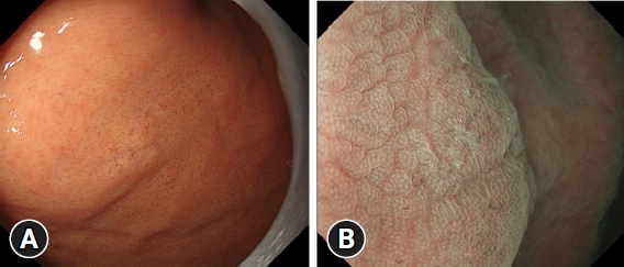
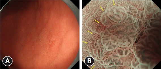
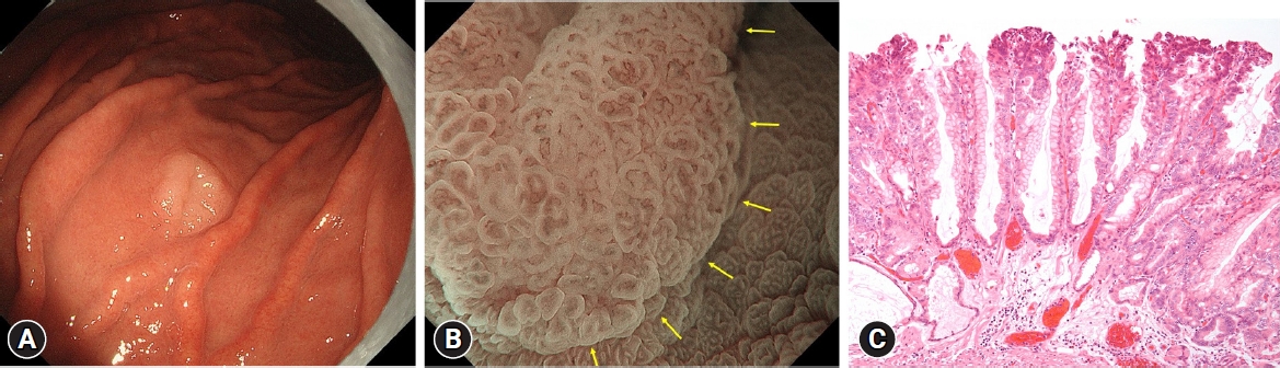
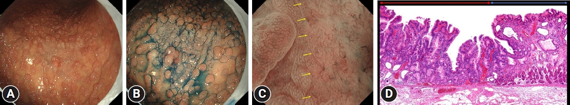
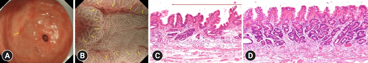
Next, HPNGC lesions were histopathologically classified (Table 2). There were 43 differentiated-type and 9 undifferentiated-type adenocarcinoma lesions. The most common differentiated type of adenocarcinoma was gastric adenocarcinoma of fundic-gland differentiation, which was observed in 30 lesions. Following further analyses of these 30 lesions, 15 were classified as adenocarcinoma of fundic-gland type (Fig. 1), 13 were classified as adenocarcinoma of fundic-gland mucosa type (Fig. 2), and 2 were categorized as adenocarcinoma of mixed fundic and pyloric-mucosa type. Additionally, 3 lesions exhibited gastric adenocarcinoma of foveolar-type, consisting of one lesion with a whitish elevation (Fig. 3) and 2 lesions presenting as raspberry-like foveolar-type gastric adenocarcinoma. Furthermore, there were 5 gastric adenocarcinoma lesions arising from polyposis, including 2 lesions observed in individuals with Peutz-Jeghers syndrome and 3 lesions observed in individuals with familial adenomatous polyposis (Fig. 4). Gastric adenocarcinoma with autoimmune gastritis was observed in one lesion. Undifferentiated-type adenocarcinoma was observed in 9 lesions, all of which were signet-ring cell carcinomas. Four well-differentiated tubular adenocarcinoma with low-grade atypia lesions were observed in the antrum (Fig. 5).
The morphology of the lesions was flat or depressed in all 9 lesions of signet-ring cell carcinoma, 3 lesions of well-differentiated tubular adenocarcinoma with low-grade atypia located in the antrum, 7 lesions of adenocarcinoma of fundic-gland type, and one lesion of adenocarcinoma of fundic-gland mucosa type (total: 20/52, 38.5%). The other lesions exhibited an elevated morphology (61.5%, 32/52).
Histopathological examination was conducted on all resected specimens, revealing the presence of gastric intestinal metaplasia in the background mucosa of 3 lesions (5.6%; 3/54 lesions). All 3 lesions were well-differentiated tubular adenocarcinomas with low-grade atypia located in the antrum.

Characteristic histological findings and common sites of HPNGC

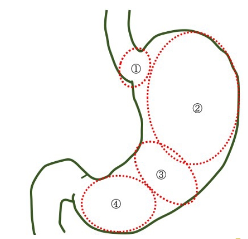
The current study revealed that the histological types and endoscopic findings of HPNGC differed among the 4 regions, as described below (Fig. 6).

1) Cardia

Advanced gastric cancer was observed in this region, with both lesions exhibiting an elevated morphology.

2) Fundic gland region ranging from the gastric fornix to the gastric body

This region comprised lesions of the following types: adenocarcinoma of fundic-gland type (Fig. 1), adenocarcinoma of fundic-gland mucosa type (Fig. 2), and adenocarcinoma of mixed fundic and pyloric-mucosa types; gastric adenocarcinoma of foveolar-type presenting with whitish elevation (Fig. 3) and raspberry-like appearance; gastric adenocarcinoma arising in polyposis, including adenocarcinoma in Peutz-Jeghers syndrome and adenocarcinoma in familial adenomatous polyposis (Fig. 4); and gastric adenocarcinoma with autoimmune gastritis.
Most lesions exhibited an elevated morphology (79.5%, 31/39).

3) Gastric-pyloric transition region ranging from the lower gastric body to the gastric angle

In this region, signet-ring cell carcinoma was observed. All lesions exhibited a flat or depressed morphology (100%, 9/9).

4) Antrum

Well-differentiated tubular adenocarcinoma with low-grade atypia (Fig. 5) was observed in this region. Most lesions exhibited a depressed morphology (75.0%, 3/4).

Diagnostic performance of M-NBI using the VS classification system for HPNGC

Three lesions that were difficult to evaluate by imaging were excluded, and 51 lesions were analyzed. The accuracy of M-NBI using the VS classification system for HPNGC diagnosis was 51.0% (26/51). No diagnosis could be made for 15 lesions of adenocarcinoma of fundic-gland type (Fig. 1B), one lesion of adenocarcinoma of fundic-gland mucosa type, 2 lesions of mixed fundic and pyloric-mucosa type, and 7 lesions of signet-ring cell carcinoma.

DISCUSSION

The current study demonstrated the clinicopathological and endoscopic features of HPNGC. The prevalence of HPNGC in the present study was 2.6% (54/2,112), which was comparable to that of 0.42% to 6.6% reported in previous studies.4-8
Herein, patients with H. pylori infection-negative advanced gastric cancer accounted for only 0.1% (2/2,112) of cases. Although HPNGC has been reported to be biologically less malignant than H. pylori infection-positive gastric cancer,13 there have been reports of advanced gastric cancer development in H. pylori infection-negative patients.14-16 The type of H. pylori infection-negative early gastric cancer that can rapidly progress to advanced gastric cancer remains unclear, which warrants further studies.
The current study revealed that tumors arising from different gastric regions exhibited distinct histological and endoscopic findings, including macroscopic types. Herein, advanced gastric cancer occurred in the cardia region. In the fundic gland region ranging from the gastric fornix to the gastric body, adenocarcinomas of fundic-gland differentiation (adenocarcinoma of fundic-gland type,17 adenocarcinoma of fundic-gland mucosa type,18,19 and adenocarcinoma of mixed fundic and pyloric-mucosa type20,21), gastric adenocarcinoma of foveolar-type (gastric adenocarcinoma of foveolar-type presenting with whitish elevation6,7 and raspberry-like foveolar-type gastric adenocarcinoma22), gastric adenocarcinoma arising in polyposis (adenocarcinoma in Peutz-Jeghers syndrome and adenocarcinoma in familial adenomatous polyposis), and gastric adenocarcinoma with autoimmune gastritis23 were observed. Signet-ring cell carcinoma was observed in the gastric-pyloric transition region ranging from the lower gastric body to the gastric angle.24,25 A well-differentiated tubular adenocarcinoma with low-grade atypia26 was observed in the antrum. Regarding the macroscopic characteristics, lesions located in the fundic gland region ranging from the gastric fornix to the gastric body were characterized by an elevated morphology, while lesions located in the gastric-pyloric transition region ranging from the lower gastric body to the gastric angle were characterized by a flat or depressed morphology. The current study revealed that the risk of advanced gastric cancer development might be present at the cardia and that it might be possible to diagnose early gastric cancer based on specific background endoscopic features. The detection of gastric cancer during endoscopic screening in patients with H. pylori infection-negative cancer may be achieved if the endoscopist is aware of the characteristic endoscopic findings and the specific localization within the stomach, which have been demonstrated in this study.
Herein, HPNGC lesions were predominantly observed in the fundic gland (72.2%, 39/54). Given that the fundic gland region has many blind spots that are hidden by greater curvature plication during the endoscopic screening of the intragastric region, extending and carefully observing the gastric wall is critical. Tumors mimic the morphology and function of their origins. Therefore, epithelial tumors arising from the H. pylori infection-negative stomach resemble cells and tissues of the proper gastric mucosa without intestinal metaplasia.27 The fundic glands are widely distributed in the gastric mucosa except for relatively small regions containing the cardiac or pyloric glands. This may explain why HPNGC was often observed in the fundic gland region in this study. In addition, it is a well-known fact that gastric adenocarcinoma of fundic-gland differentiation arises from the mucosa in the fundic gland region. The inclusion of several patients with this type of tumor in this study may be an additional reason for the frequent observation of HPNGC within the fundic gland mucosa. The current study confirmed gastric intestinal metaplasia, even in background mucosa without H. pylori infection, in 3 lesions, all located in the antrum. Recently, bile reflux has been linked to gastric intestinal metaplasia.28-31 The development of intestinal metaplasia may be attributable to the predisposition of the antrum to bile reflux.
M-NBI showed that 27 lesions (53%) of HPNGC met the diagnostic criteria for cancer according to the VS classification system, and no diagnosis could be made for 15 lesions of adenocarcinoma of fundic-gland type, one lesion of adenocarcinoma of fundic-gland mucosa type, 2 lesions of adenocarcinoma of mixed fundic and pyloric-mucosa type, and 7 lesions of signet-ring cell carcinoma. A histological architecture that makes diagnosis using M-NBI difficult is the superficial layer of the intramucosal tumor tissue being widely covered by non-tumor epithelium, as seen in adenocarcinoma of the fundic-gland type and some undifferentiated-type adenocarcinomas. Such lesions require the histological examination of biopsy specimens for definite diagnosis.
The limitation of the current study is its single-center retrospective design.
In conclusion, the study findings demonstrated that HPNGC originating from various regions of the stomach exhibited distinct endoscopic characteristics and histological types. These specific features related to cancer sites should be considered when performing screening endoscopies for individuals with H. pylori infection-negative stomach to enhance diagnostic accuracy and effectiveness.

NOTES

Conflicts of Interest
Kenshi Yao is a member of the editorial board of Clinical Endoscopy; however, he was not involved in peer reviewer selection, evaluation, or the decision process in this study. The other authors have no potential conflicts of interest.
Funding
None.
Author Contributions
Conceptualization: all authors; Data curation: KI; Formal analysis: KI; Investigation: KI; Methodology: KI, KY; Project administration: KI, KY; Resources: KI; Supervision: all authors; Visualization: KI; Writing–original draft: KI; Writing–review & editing: all authors.

Fig. 1.
Endoscopic findings of gastric adenocarcinoma of fundic-gland type. (A) Conventional endoscopic image: a pale subepithelial tumor-like elevated lesion is observed in the fundus. Dilated vessels can be seen in the elevated region. (B) Image of the lesion obtained using magnifying endoscopy with narrow-band imaging. Upon observing the surrounding area, the lesion shows no marked changes in the microvascular (MV) or microsurface (MS) pattern, and there is no clear demarcation line (DL). The microvessel has a polygonal closed-loop morphology, and the vessels are uniform in shape, arranged regularly, and symmetrically distributed. The marginal crypt epithelium has an oval or arc-shaped morphology, is uniform in shape, arranged regularly, and distributed symmetrically. According to the vessel plus surface classification system, the lesion is classified as having a regular MV pattern plus a regular MS pattern without DL. In other words, the magnifying endoscopic findings of the lesion does not fulfill the diagnostic criteria for cancer.
ce-2023-258f1.jpg
Fig. 2.
Endoscopic findings of gastric adenocarcinoma of fundic-gland mucosa type. (A) Conventional endoscopic image. The lesion has a slightly red mucosa in the posterior wall of the lower gastric body. (B) Image of the lesion obtained using magnifying endoscopy with narrow-band imaging. Upon observing the background mucosa, the elevated lesion shows marked changes in both the microvascular (MV) and microsurface (MS) patterns, and there is a clear demarcation line (DL) (yellow arrows). Regarding the MV pattern, the lesion consists of microvessels with open- and closed-loop formation, and the vessels are not uniform in shape, distributed asymmetrically, and arranged irregularly. Regarding the MS pattern, the marginal crypt epithelium (MCE) has a curved or oval morphology. The MCE is nonuniform in shape, distributed asymmetrically, and arranged irregularly. According to the vessel plus surface classification system, the lesion was classified as having an irregular MV pattern plus an irregular MS pattern with a DL, and was diagnosed as cancer.
ce-2023-258f2.jpg
Fig. 3.
Endoscopic and histopathological findings of gastric adenocarcinoma of foveolar-type presenting with whitish elevation. (A) Conventional endoscopic findings. A markedly whitish elevated lesion is observed in the greater curvature of the upper gastric body. (B) Image of the lesion obtained using magnifying endoscopy with narrow-band imaging. Upon observation of the background mucosa, the lesion shows marked changes both in microvascular (MV) and microsurface (MS) patterns, and there is a clear demarcation line (DL) (yellow arrows). With respect to the MV pattern, the lesion consists of microvessels with open- and closed-loop formation, and the vessels are not uniform in shape, distributed asymmetrically, and arranged irregularly. With respect to the MS pattern, the marginal crypt epithelium (MCE) has a curved or oval morphology. The MCE is nonuniform in shape, distributed asymmetrically, and arranged irregularly. According to the vessel plus surface classification system, the lesion was classified as having an irregular MV pattern plus an irregular MS pattern with a DL, and was diagnosed as cancer. (C) Histopathological image of gastric adenocarcinoma of foveolar-type presenting with whitish elevation (hematoxylin and eosin stain, ×100). Tall cells with clear cytoplasm are proliferating in a papillary pattern. They exhibit histopathological features of foveolar-type gastric adenocarcinoma.
ce-2023-258f3.jpg
Fig. 4.
Endoscopic and histopathological findings of adenocarcinoma in familial adenomatous polyposis. (A) Conventional endoscopic image. Multiple fundic gland polyps can be seen developing in a region ranging from the gastric fornix to the gastric body. A whitish area is observed in the greater curvature of the upper gastric body. (B) Imaging result of indigocarmine staining. The whitish area appears to have a smaller granular elevation than the surrounding area. (C) Image of the lesion obtained using magnifying endoscopy with narrow-band imaging. Upon observation of the background mucosa, the lesion shows marked changes both in the microvascular (MV) and microsurface (MS) patterns, and there is a clear demarcation line (DL) (yellow arrows). With respect to the MV pattern, the lesion consists of microvessels with open- and closed- loop formation, and the vessels are not uniform in shape, distributed asymmetrically, and arranged irregularly. With respect to the MS pattern, almost no marginal crypt epithelium can be visualized. According to the vessel pulse surface classification system, the lesion was classified as having an irregular MV pattern plus an absent MS pattern with a DL, and was diagnosed as cancer. (D) Histopathological image of adenocarcinoma in familial adenomatous polyposis (hematoxylin and eosin stain, ×40). The histological findings of well-differentiated tubular adenocarcinoma are observed (red line). The mucosa surrounding the tumor exhibits the findings of fundic gland polyps with fundic gland hyperplasia and cyst formation (blue line).
ce-2023-258f4.jpg
Fig. 5.
Endoscopic and histopathological findings of well-differentiated adenocarcinoma with low-grade atypia. (A) Conventional endoscopic image. There are multiple small red depressed lesions in the antrum. The yellow arrow indicates the lesion. (B) Image of the lesion obtained using magnifying endoscopy with narrow-band imaging. The lesion shows marked changes in both microvascular (MV) and microsurface (MS) patterns, with a clear demarcation line (DL) (yellow arrows). With respect to the MV pattern, the lesion mainly consists of microvessels with open-loop formation, and the vessels are not uniform in shape, distributed asymmetrically, and arranged irregularly. With respect to the MS pattern, the marginal crypt epithelium (MCE) has a curved morphology. The MCE is nonuniform in shape, distributed asymmetrically, and arranged irregularly. According to the vessel plus surface classification system, the lesion was classified as having an irregular MV pattern plus an irregular MS pattern with a DL, and was diagnosed as cancer. (C) Histopathological appearance (hematoxylin and eosin stain, ×40). The tumor shows slightly irregular branching ducts with mildly enlarged nuclei and is diagnosed as a well-differentiated adenocarcinoma with low-grade atypia (red arrow). (D) Histopathological findings in the background mucosa of the lesion (hematoxylin and eosin stain, ×40). Gastric intestinal metaplasia is observed.
ce-2023-258f5.jpg
Fig. 6.
Characteristics of the preferential development sites of Helicobacter pylori infection-negative gastric cancer. (1) Cardia: advanced gastric cancer. (2) Fundic gland region ranging from the gastric fornix to the gastric body: adenocarcinoma of fundic gland differentiation (adenocarcinoma of fundic-gland type, adenocarcinoma of fundic-gland mucosa type, adenocarcinoma of mixed fundic and pyloric-mucosa types), gastric adenocarcinoma of foveolar-type (gastric adenocarcinoma of foveolar-type presenting with whitish elevation, raspberry-like foveolar-type gastric adenocarcinoma), gastric adenocarcinoma arising in polyposis (adenocarcinoma in Peutz-Jeghers syndrome, adenocarcinoma in familial adenomatous polyposis), and gastric adenocarcinoma with autoimmune gastritis. (3) Gastric-pyloric transition region ranging from the lower gastric body to the gastric angle: signet-ring cell carcinoma. (4) Antrum: well-differentiated adenocarcinoma with low-grade atypia.
ce-2023-258f6.jpg
ce-2023-258f7.jpg
Table 1.
Clinicopathological features of Helicobacter pylori infection-negative gastric cancer (n=54)
Value
Mean age (yr) 61.1 (19–82)
Sex
 Male 26 (48.1)
 Female 28 (51.9)
Macroscopic type
 Elevated 34 (63.0)
 Flat or depressed 20 (37.0)
Depth of invasion
 Mucosa 33 (61.1)
 Submucosa 19 (35.2)
 Muscularis propria or subserosa 2 (3.7)
Lymphovascular invasion
 Positive 2 (3.7)

Values are presented as mean (range) or number (%).

Table 2.
Classification of pathological histology of Helicobacter pylori infection-negative early gastric cancer (n=52)
Value
Differentiated-type adenocarcinoma
 Gastric adenocarcinoma of fundic-gland differentiation
  Adenocarcinoma of fundic-gland type 15
  Adenocarcinoma of fundic-gland mucosa type 13
  Adenocarcinoma of mixed fundic and pyloric-mucosa type 2
 Gastric adenocarcinoma of foveolar-type
  Gastric adenocarcinoma of foveolar-type presenting with whitish elevation 1
  Raspberry-like foveolar-type gastric adenocarcinoma 2
 Gastric adenocarcinoma arising in polyposis
  Adenocarcinoma in Peutz-Jeghers syndrome 2
  Adenocarcinoma in familial adenomatous polyposis 3
 Gastric adenocarcinoma with autoimmune gastritis 1
 Well-differentiated tubular adenocarcinoma with low-grade atypia 4
Undifferentiated-type adenocarcinoma
 Signet-ring cell carcinoma 9

REFERENCES

1. Warren JR, Marshall B. Unidentified curved bacilli on gastric epithelium in active chronic gastritis. Lancet 1983;1:1273–1275.
crossref pmid
2. Infection with Helicobacter pylori. IARC Monogr Eval Carcinog Risks Hum 1994;61:177–240.
pmid pmc
3. Kamada T, Haruma K, Ito M, et al. Time trends in Helicobacter pylori infection and atrophic gastritis over 40 years in Japan. Helicobacter 2015;20:192–198.
crossref pmid pmc
4. Ono S, Kato M, Suzuki M, et al. Frequency of Helicobacter pylori -negative gastric cancer and gastric mucosal atrophy in a Japanese endoscopic submucosal dissection series including histological, endoscopic and serological atrophy. Digestion 2012;86:59–65.
crossref pmid pdf
5. Kim HJ, Kim N, Yoon H, et al. Comparison between resectable Helicobacter pylori-negative and -positive gastric cancers. Gut Liver 2016;10:212–219.
crossref pmid
6. Yamada A, Kaise M, Inoshita N, et al. Characterization of Helicobacter pylori-naïve early gastric cancers. Digestion 2018;98:127–134.
crossref pmid pdf
7. Sato C, Hirasawa K, Tateishi Y, et al. Clinicopathological features of early gastric cancers arising in Helicobacter pylori uninfected patients. World J Gastroenterol 2020;26:2618–2631.
crossref pmid pmc
8. Akazawa Y, Ueyama H, Hayashi T, et al. Clinicopathological and molecular characterization of early gastric adenocarcinoma in Helicobacter pylori-uninfected patients: emphasis on differentiated gastric adenocarcinoma. J Gastroenterol 2022;57:725–734.
crossref pmid pdf
9. Kimura K, Takemoto T. An endoscopic recognition of the atrophic border and its significance in chronic gastritis. Endoscopy 1969;1:87–97.
crossref
10. Sakaki N, Momma K, Egawa N, Yamada Y, Kan T, Ishiwata J. The influence of Helicobacter pylori infection on the progression of gastric mucosal atrophy and occurrence of gastric cancer. Eur J Gastroenterol Hepatol 1995;7 Suppl 1:S59–S62.
pmid
11. Yao K. The endoscopic diagnosis of early gastric cancer. Ann Gastroenterol 2013;26:11–22.
pmid pmc
12. Yao K, Anagnostopoulos GK, Ragunath K. Magnifying endoscopy for diagnosing and delineating early gastric cancer. Endoscopy 2009;41:462–467.
crossref pmid
13. Yoon H, Kim N, Lee HS, et al. Helicobacter pylori-negative gastric cancer in South Korea: incidence and clinicopathologic characteristics. Helicobacter 2011;16:382–388.
crossref pmid
14. Okano A, Kato S, Ohana M. Helicobacter pylori-negative gastric cancer: advanced-stage undifferentiated adenocarcinoma located in the pyloric gland area. Clin J Gastroenterol 2017;10:13–17.
crossref pmid pdf
15. Hayashi J, Yamatsuji T, Suehiro M, et al. Helicobacter pylori-negative advanced gastric cancer arising from the gastric mucosa without inflammation, atrophy, or intestinal metaplasia. Case Rep Gastroenterol 2022;16:345–350.
crossref pmid pmc pdf
16. Takagi A, Ozawa H, Oki M, Yanagi H, Nabeshima K, Nakamura N. Helicobacter pylori-negative advanced gastric cancer with massive eosinophilia. Intern Med 2018;57:1715–1718.
crossref pmid pmc
17. Ueyama H, Yao T, Nakashima Y, et al. Gastric adenocarcinoma of fundic gland type (chief cell predominant type): proposal for a new entity of gastric adenocarcinoma. Am J Surg Pathol 2010;34:609–619.
crossref pmid
18. Ueyama H, Yao T, Akazawa Y, et al. Gastric epithelial neoplasm of fundic-gland mucosa lineage: proposal for a new classification in association with gastric adenocarcinoma of fundic-gland type. J Gastroenterol 2021;56:814–828.
crossref pmid pmc pdf
19. Imamura K, Yao K, Nimura S, et al. Characteristic endoscopic findings of gastric adenocarcinoma of fundic-gland mucosa type. Gastric Cancer 2021;24:1307–1319.
crossref pmid pdf
20. Kanesaka T, Uedo N, Yao K, et al. New subtype of gastric adenocarcinoma: mixed fundic and pyloric mucosa-type adenocarcinoma. Clin J Gastroenterol 2017;10:224–228.
crossref pmid pdf
21. Takahashi H, Yao K, Ueo T, et al. Histological subtype of gastric adenocarcinoma: two cases of mixed fundic and pyloric mucosa-type adenocarcinoma. Ecancermedicalscience 2020;14:1143.
crossref pmid pmc
22. Shibagaki K, Fukuyama C, Mikami H, et al. Gastric foveolar-type adenomas endoscopically showing a raspberry-like appearance in the Helicobacter pylori-uninfected stomach. Endosc Int Open 2019;7:E784–E791.
crossref pmid pmc
23. Arai J, Niikura R, Hayakawa Y, et al. Clinicopathological features of gastric cancer with autoimmune gastritis. Biomedicines 2022;10:884.
crossref pmid pmc
24. Nikaido M, Kakiuchi N, Miyamoto S, et al. Indolent feature of Helicobacter pylori-uninfected intramucosal signet ring cell carcinomas with CDH1 mutations. Gastric Cancer 2021;24:1102–1114.
crossref pmid pdf
25. Muraishi J, Miyaoka M, Imamura K, et al. A case of gastric signet-ring cell carcinoma with a long-term retrospective follow-up of 17 years. Clin J Gastroenterol 2021;14:1337–1343.
crossref pmid pdf
26. Takita M, Ohata K, Inamoto R, et al. Endoscopic and histological features of Helicobacter pylori-negative differentiated gastric adenocarcinoma arising in the antrum. JGH Open 2021;5:470–477.
crossref pmid pmc pdf
27. Kushima R, Nimura S. Stomach. In: Fukayama M, Morinaga S, editors. Surgical pathology. 5th ed. Bunkodo; 2020. p. 444–515.

28. Matsuhisa T, Tsukui T. Relation between reflux of bile acids into the stomach and gastric mucosal atrophy, intestinal metaplasia in biopsy specimens. J Clin Biochem Nutr 2012;50:217–221.
crossref pmid
29. Tatsugami M, Ito M, Tanaka S, et al. Bile acid promotes intestinal metaplasia and gastric carcinogenesis. Cancer Epidemiol Biomarkers Prev 2012;21:2101–2107.
crossref pmid pdf
30. Matsuhisa T, Arakawa T, Watanabe T, et al. Relation between bile acid reflux into the stomach and the risk of atrophic gastritis and intestinal metaplasia: a multicenter study of 2283 cases. Dig Endosc 2013;25:519–525.
crossref pmid
31. Li D, Zhang J, Yao WZ, et al. The relationship between gastric cancer, its precancerous lesions and bile reflux: a retrospective study. J Dig Dis 2020;21:222–229.
crossref pmid pmc pdf
TOOLS
PDF Links  PDF Links
PubReader  PubReader
ePub Link  ePub Link
XML Download  XML Download
Full text via DOI  Full text via DOI
Download Citation  Download Citation
  Print
Share:      
METRICS
0
Crossref
0
Scopus
2,013
View
32
Download
Related articles
Improving the quality of the esophagogastroduodenoscopy in Helicobacter pylori-naïve gastric cancer  2023 July;56(4)
Exploring quality indicators for the detection of Helicobacter pylori-naïve gastric cancer: a cross-sectional nationwide survey  2023 July;56(4)
Quality Indicators for the Detection of Helicobacter pylori-Negative Early Gastric Cancer: A Retrospective Observational Study  2020 November;53(6)
Editorial Office
Korean Society of Gastrointestinal Endoscopy
#817, 156 Yanghwa-ro (LG Palace, Donggyo-dong), Mapo-gu, Seoul, 04050, Korea
TEL: +82-2-335-1552   FAX: +82-2-335-2690    E-mail: CE@gie.or.kr
Copyright © Korean Society of Gastrointestinal Endoscopy.                 Developed in M2PI
Close layer