Clin Endosc > Volume 57(2); 2024 > Article
Hara, Okuno, Haba, and Kuwahara: Forward viewing liner echoendoscopy for therapeutic interventions

Abstract

Therapeutic endoscopic ultrasonography (EUS) procedures using the forward-viewing convex EUS (FV-EUS) have been reviewed based on the articles reported to date. The earliest reported procedure is the drainage of pancreatic pseudocysts using FV-EUS. However, the study on drainage of pancreatic pseudocysts focused on showing that drainage is possible with FV-EUS rather than leveraging its features. Subsequently, studies describing the characteristics of FV-EUS have been reported. By using FV-EUS in EUS-guided choledochoduodenostomy, double punctures in the gastrointestinal tract can be avoided. In postoperative modified anatomical cases, using the endoscopic function of FV-EUS, procedures such as bile duct drainage from anastomosis, pancreatic duct drainage from the afferent limb, and abscess drainage from the digestive tract have been reported. When a perpendicular puncture to the gastrointestinal tract is required or when there is a need to insert the endoscope deep into the gastrointestinal tract, FV-EUS is considered among the options.

INTRODUCTION

Endoscopic ultrasonography (EUS) has become an essential technique for the diagnosis of biliary and pancreatic diseases and is widely adopted worldwide. Recently, the use of EUS has expanded beyond just biliary and pancreatic diseases to allow detailed observation of the gastrointestinal tract and its adjacent organs. Procedures such as EUS-guided fine-needle aspiration (EUS-FNA) are being conducted, and genetic diagnoses based on this method are being performed. EUS has evolved from being merely diagnostic to therapeutic EUS. Although the oblique-viewing convex EUS (OV-EUS) is commonly used for therapeutic purposes, using the forward-viewing convex EUS (FV-EUS) can improve safety and overcome further challenges. In this review, we discuss the results of therapeutic EUS using the FV-EUS.

PROS AND CONS IN FV-EUS

FV-EUS is an endoscopy technique that combines standard forward-viewing endoscopy with ultrasound imaging, allowing the physician to view the digestive tract and obtain high-quality ultrasound images of organs and lesions within or adjacent to the digestive tract. The forward-view allows for easy navigation through the gastrointestinal tract, which is especially beneficial for patients with surgically altered anatomy and accessing certain anatomical sites. FV-EUS also allows for vertical puncture in the gastrointestinal tract, leading to improved puncture performance.1 FV-EUS is highly useful for approaching areas that are difficult to reach with OV-EUS and when wanting to puncture the gastrointestinal tract vertically as shown in Figure 1 and Table 1. Cons of FV-EUS include its EUS imaging range being limited to 90° and the absence of an elevator, which makes adjusting the puncture angle challenging.

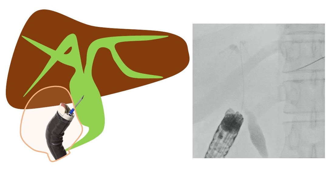
EUS-GUIDED CHOLEDOCHODUODENOSTOMY

Hara et al. have reported a prospective study using FV-EUS for primary EUS-guided choledochoduodenostomy (EUS-CDS).2 This study describes the first prospective trial on primary EUS-guided biliary drainage and is the first study to examine the utility of FV-EUS in EUS-CDS. The technical and functional success rates were 94.4% (17/18) and 94.1% (16/17), respectively. Early adverse events (focal peritonitis) were encountered in 2 patients (11.8%). No patients developed late adverse events. These results are highly promising. Matsumoto et al.3 have reported that by using FV-EUS during EUS-CDS, double puncture of the digestive tract can be prevented. In OV-EUS, the axis of the scope and the puncture needle is different, resulting in the needle being inserted obliquely into the digestive tract, leading to a double puncture of the mucosa or difficulty in transmitting force.4 However, by using FV-EUS, these challenges can be overcome (Figs. 1, 2). When placing a stent, endoscopic imaging is easily available, allowing for the stent to be safely positioned in the appropriate position.1-3,5-7 For similar reasons, FV-EUS may also be useful for EUS-gallbladder drainage (EUS-GBD). It is believed that, by using FV-EUS, it is possible to avoid transgastric puncture and double puncture of the digestive tract mucosa in EUS-CDS and EUS-GBD. There are no reports summarizing the clinical outcomes of EUS-GBD.

EUS-GUIDED HEPATICOGASTROSTOMY

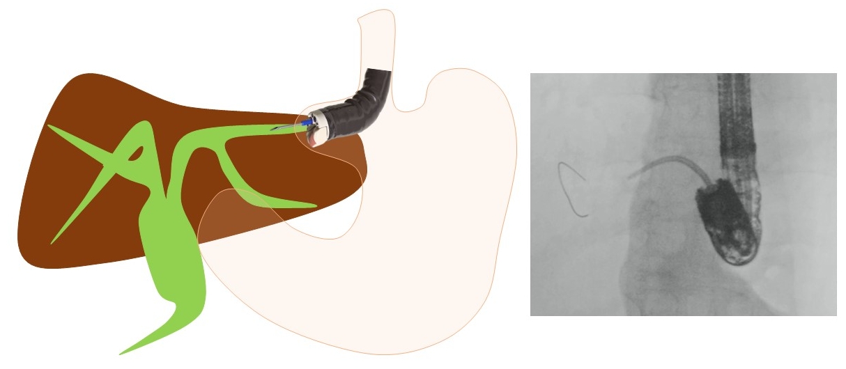
When performing EUS-guided hepaticogastrostomy (EUS-HGS), Hara and Okuno et al.8,9 first reported that, by using FV-EUS, a 22-G needle, and an 018 wire (Fig. 3), B2 puncture is available and the procedure becomes easier. When carrying out EUS-HGS, it is common to perform a B3 puncture to avoid transesophageal puncture. However, choosing B2 may be more convenient for tasks such as inserting the guidewire and some devices. If B2 can be punctured safely, it makes the procedure easier. According to a report by Okuno et al.,9 B2-EUS-HGS using FV-EUS has a success rate of 88.5% (54/61), with a complication rate of only 6.5% (4/61). There were no instances of transesophageal puncture. Moreover, Elshair et al.10 have reported the potential utility of techniques using FV-EUS when placing HGS in both B2 and B3.

EUS-GUIDED HEPATICOJEJUNOSTOMY

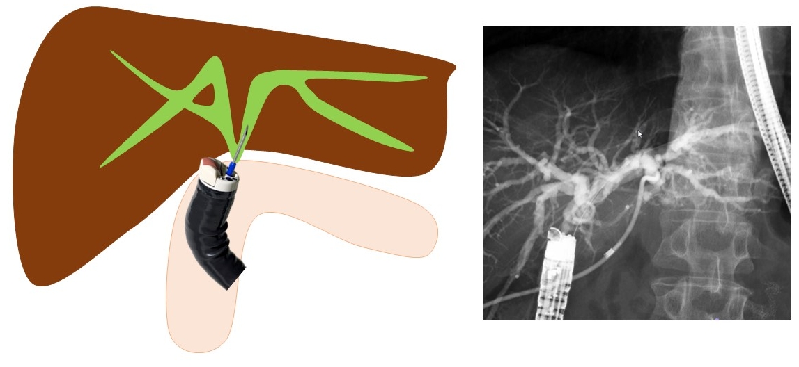
Kida et al.11 have first reported that in cases of EUS-guided hepaticojejunostomy (EUS-HJS), the bile duct is punctured using FV-EUS from the jejunum of the anastomosis, and a stent is placed (Fig. 4). Because the puncture is performed via the anastomosis, the risk of bile leakage is low, and drainage can be performed relatively safely. Although EUS-HGS can also treat anastomosis stenosis, it is difficult to drain the right side in cases where the left and right are separated. However, using this method, it is possible to drain both left and right hepatic ducts via the anastomosis. If a stent can be placed through the anastomosis, there is a possibility that the stent can ultimately be removed and made stent-free, and thus it is considered a first-line treatment method, especially in benign stenosis cases. Ishiwatari et al.12 reported that, in recurrent cases after pancreaticoduodenectomy, they performed EUS-HJS using FV-EUS from the afferent limb to drain the right bile duct of the liver. The limitation of this technique is the difficulty in inserting the FV-EUS up to the bile duct-jejunum anastomosis. These reports on EUS-HJS using FV-EUS are limited to case reports,11-20 and there are no published studies summarizing the clinical results. It is hoped that more extensive studies and reports will be conducted in the future.

EUS-GUIDED PANCREATICOJEJUNOSTOMY

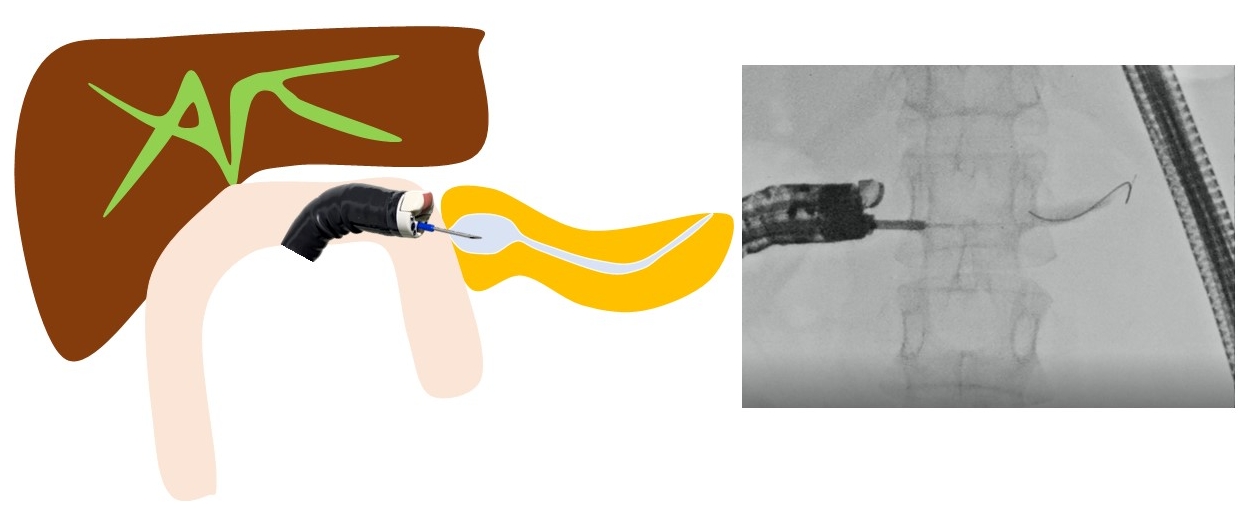
Endoscopic treatment for pancreatic duct-jejunum anastomosis stenosis is more challenging and prone to failure than that for bile duct-jejunum anastomosis stenosis. The gold standard is to dilate the anastomosis using a balloon endoscope and place a stent. However, if the usual method is challenging, treatment under EUS is effective. When using OV-EUS, it is common to approach from the stomach.21 However, there is no adhesion between the pancreas and stomach, which raises issues such as stent migration and misplacement, pancreatic fluid leakage, and pancreatitis. To address these problems, Nakaji et al.22 have reported on EUS-guided pancreaticojejunostomy (EUS-PJS). EUS-PJS is a technique that involves placing a pancreatic duct stent via the anastomosis and through the small intestine.11,21-27 Because there is a lower risk of postoperative stent misplacement into the abdominal cavity or pancreatic fluid leakage, it can be safely performed (Fig. 5). When inserting the stent via the stomach, in some cases, the guidewire cannot penetrate the anastomosis, necessitating permanent stent replacement. However, if approached through the anastomosis, there is a possibility to make it stent-free. These reports on EUS-PJS using FV-EUS have been limited to case reports,22-27 and there are no published studies summarizing the clinical results. More comprehensive studies and reports are anticipated in the future.

EUS-GUIDED PANCREATIC CYST DRAINAGE

EUS-guided pancreatic cyst drainage using EUS-FV was first reported by Voermans et al.28 It is believed that there are advantages to utilizing the endoscopic capabilities characteristic of FV. It is possible to treat digestive tract stenosis, visualize the stent after placement, and other options.1,7,29-32 However, because there is no difference in the technical and clinical success rate of this technique, the benefits of using FV-EUS seem to be limited.

OTHER TREATMENTS

Other reported treatments include drainage for intra-abdominal abscess (Fig. 6), liver abscess, splenic abscess, and endovascular treatments for gastric varices.33-38 FV-EUS may be used if it is possible to insert the endoscope deeply into the digestive tract, if puncturing perpendicularly to the digestive tract is feasible, and if endoscopic images are useful for the procedures.

CONCLUSIONS

FV-EUS presents a significant advancement in the field of gastrointestinal endoscopy, combining the advantages of both standard endoscopy and ultrasound imaging. Although it may not entirely replace traditional OV-EUS owing to certain limitations, its specific applications and advantages make it a valuable tool in both diagnostic and therapeutic endoscopy.

NOTES

Conflicts of Interest
The authors have no potential conflicts of interest.
Funding
None.
Author Contributions
Conceptualization: all authors; Project administration: all authors; Supervision: all authors; Writing–original draft: KH, SH, TK; Writing–review & editing: KH, NO.

Fig. 1.
Difference between forward-viewing convex endoscopic ultrasonography (FV-EUS) and oblique-viewing convex EUS (OV-EUS). The difference in puncture angles between FV-EUS and OV-EUS is shown. FV-EUS allows for a vertical puncture relative to the digestive tract, whereas OV-EUS is punctured at an angle to the digestive tract.
ce-2023-271f1.jpg
Fig. 2.
Endoscopic ultrasonography (EUS)-guided choledochoduodenostomy. The image shows the extrahepatic bile duct being punctured using forward-viewing convex EUS. A vertical puncture relative to the digestive tract is possible, avoiding double punctures of the digestive tract.
ce-2023-271f2.jpg
Fig. 3.
Endoscopic ultrasonography (EUS)-guided hepaticogastrostomy. The image depicts B2 being punctured with forward-viewing convex EUS (FV-EUS) and the fistula expanded using a drill dilator. Using FV-EUS makes puncturing B2 easier and safer.
ce-2023-271f3.jpg
Fig. 4.
Endoscopic ultrasonography-guided hepaticojejunostomy. A case of bile duct-jejunum anastomosis stenosis after a pancreaticoduodenectomy. From the afferent limb, anastomosis is punctured to place a stent.
ce-2023-271f4.jpg
Fig. 5.
Endoscopic ultrasonography-guided pancreaticojejunostomy. A case of pancreatic duct-jejunum anastomosis stenosis after a pancreaticoduodenectomy. By puncturing from the anastomosis side, the risk of pancreatic juice leakage and other complications is reduced.
ce-2023-271f5.jpg
Fig. 6.
Endoscopic ultrasonography (EUS)-abscess drainage from the afferent limb. For draining areas such as liver abscesses on the dorsal side of the liver and intraperitoneal abscesses where percutaneous drainage is difficult, drainage using forward-viewing convex EUS is effective.
ce-2023-271f6.jpg
Table 1.
Pros and cons in forward-viewing convex endoscopic ultrasonography
Pros Cons
Forward endoscopic view No elevator
Easy scope insertion Narrow scanning range
Vertical puncture to the gastrointestinal tract
Preventing double puncture of the mucosa
Wide angulation angle

REFERENCES

1. Kida M, Araki M, Miyazawa S, et al. Fine needle aspiration using forward-viewing endoscopic ultrasonography. Endoscopy 2011;43:796–801.
crossref pmid
2. Hara K, Yamao K, Hijioka S, et al. Prospective clinical study of endoscopic ultrasound-guided choledochoduodenostomy with direct metallic stent placement using a forward-viewing echoendoscope. Endoscopy 2013;45:392–396.
crossref pmid
3. Matsumoto S, Hara K, Mizuno N, et al. Risk factor analysis for adverse events and stent dysfunction of endoscopic ultrasound-guided choledochoduodenostomy. Dig Endosc 2020;32:957–966.
crossref pmid pdf
4. Hara K, Yamao K, Mizuno N, et al. Endoscopic ultrasonography-guided biliary drainage: who, when, which, and how? World J Gastroenterol 2016;22:1297–1303.
crossref pmid pmc
5. Ishikawa S, Okuno N, Hara K, et al. Refractory benign biliary stricture due to chronic pancreatitis in two patients treated using endoscopic ultrasound-guided choledochoduodenostomy fistula creation: case reports. Clin Endosc 2024;57:122–127.
crossref
6. Kuraoka N, Hara K, Okuno N, et al. Outcomes of EUS-guided choledochoduodenostomy as primary drainage for distal biliary obstruction with covered self-expandable metallic stents. Endosc Int Open 2020;8:E861–E868.
crossref pmid pmc
7. Trevino JM, Varadarajulu S. Initial experience with the prototype forward-viewing echoendoscope for therapeutic interventions other than pancreatic pseudocyst drainage (with videos). Gastrointest Endosc 2009;69:361–365.
crossref pmid
8. Hara K, Okuno N, Haba S, et al. How to perform EUS-guided hepaticogastrostomy easier and safer. J Hepatobiliary Pancreat Sci 2020;27:563–564.
crossref pmid pdf
9. Okuno N, Hara K, Mizuno N, et al. B2 puncture with forward-viewing EUS simplifies EUS-guided hepaticogastrostomy (with video). Endosc Ultrasound 2022;11:319–324.
crossref pmid pmc
10. Elshair M, Hara K, Haba S, et al. Combined duodenal stenting and endoscopic ultrasound-guided hepaticogastrostomy using forward-viewing echoendoscope: a one-scope technique. Endoscopy 2022;54:E516–E517.
crossref pmid
11. Kida M, Yamauchi H, Okuwaki K, et al. Endoscopic ultrasound-guided choledochojejunostomy with a forward-viewing echoendoscope for severe benign bilioenteric stricture in a patient with Child's resection. Endoscopy 2015;47 Suppl 1 UCTN:E303–E304.
crossref pmid
12. Ishiwatari H, Sato J, Kaneko J. Hepaticojejunostomy for the right hepatic bile duct using a forward-viewing echoendoscope in a patient after pancreatoduodenectomy. Dig Endosc 2019;31:e82–e83.
crossref pmid pdf
13. Kawasaki Y, Hijioka S, Takeshita K, et al. Endoscopic ultrasound-guided choledochojejunostomy using a forward-viewing echoendoscopic saddle-cross technique. Endoscopy 2023;55:E233–E235.
crossref pmid
14. Nakatsubo R, Yamamoto K, Itoi T. Endoscopic ultrasound-guided biliary drainage for complete obstruction of hepaticojejunostomy using a forward-viewing echoendoscope and novel spiral dilator. Dig Endosc 2022;34:e162–e163.
crossref pmid pdf
15. Koizumi K, Masuda S, Shionoya K, et al. Endoscopic ultrasound-guided hepaticojejunostomy using forward-viewing echoendoscope for transected aberrant right posterior hepatic duct in Roux-en-Y hepaticojejunostomy. Endoscopy 2022;54:E933–E934.
crossref pmid pmc
16. Iwai T, Kida M, Yamauchi H, et al. EUS-guided transanastomotic drainage for severe biliopancreatic anastomotic stricture using a forward-viewing echoendoscope in patients with surgically altered anatomy. Endosc Ultrasound 2021;10:33–38.
crossref pmid pmc
17. Hisada Y, Hijioka S, Ohba A, et al. Novel endoscopic ultrasound-guided hepaticoduodenostomy using a forward-viewing echoendoscope for altered anatomy. Endoscopy 2021;53:E340–E342.
crossref pmid
18. Shimizu T, Sato K, Abe T, et al. Forward-viewing echoendoscope is useful for recanalization of postoperative biliary anastomotic atresia in endosonography-guided biliary drainage. Endoscopy 2020;52:E437–E438.
crossref pmid
19. Kin T, Hayashi T, Katanuma A. Endoscopic ultrasound-guided fistulation between bile duct and afferent limb for treatment of complete choledochojejunal obstruction using forward-viewing echoendoscope. Dig Endosc 2019;31:e97–e98.
crossref pmid pdf
20. Fuccio L, Attili F, Larghi A. Forward-viewing linear echoendoscope: a new option in the endoscopic ultrasound armamentarium (with video). J Hepatobiliary Pancreat Sci 2015;22:27–34.
crossref pmid
21. Teh JL, Teoh AY. Techniques and outcomes of endoscopic ultrasound guided-pancreatic duct drainage (EUS-PDD). J Clin Med 2023;12:1626.
crossref pmid pmc
22. Nakaji S, Hirata N, Shiratori T, et al. Endoscopic ultrasound-guided pancreaticojejunostomy with a forward-viewing echoendoscope as a treatment for stenotic pancreaticojejunal anastomosis. Endoscopy 2015;47 Suppl 1 UCTN:E41–E42.
crossref pmid
23. Shimizu H, Suzuki R, Sato Y, et al. Transjejunal endoscopic ultrasound-guided pancreatic drainage for pancreatic jejunostomy stricture using a forward-viewing echoendoscope in a patient with altered anatomy. DEN Open 2022;2:e114.
crossref pmid pmc pdf
24. Nakamura K, Takigawa Y, Masuda Y, et al. Forward-viewing echoendoscope-guided pancreatic duct drainage using a diathermic dilator for a severely stenotic pancreaticojejunal anastomosis. Endoscopy 2022;54:E75–E76.
crossref pmid
25. Yoshimura S, Nakaji S, Shiratori T, et al. Endoscopic ultrasound-guided pancreaticoduodenostomy with a forward-viewing echoendoscope. VideoGIE 2021;6:549–551.
crossref pmid pmc
26. Kuwatani M, Kato S, Sakamoto N. Direct recanalization of pancreaticogastrostomy obstruction with a forward-viewing echoendoscope. Dig Endosc 2019;31:e18–e19.
crossref pmid pdf
27. Hodo Y, Shirota Y, Suda T, et al. Successful EUS-guided retrograde pancreatic duct stent placement for refractory pancreaticojejunostomy stricture after pancreaticoduodenectomy with a forward-viewing echoendoscope. VideoGIE 2018;3:196–198.
crossref pmid pmc
28. Voermans RP, Eisendrath P, Bruno MJ, et al. Initial evaluation of a novel prototype forward-viewing US endoscope in transmural drainage of pancreatic pseudocysts (with videos). Gastrointest Endosc 2007;66:1013–1017.
crossref pmid
29. Rana SS, Lal SB. Esophageal obstruction in a child secondary to large walled-off necrosis: rescue transmural drainage using forward-viewing echoendoscope. Ann Gastroenterol 2021;34:123.
crossref pmid
30. Fernández de Castro C, Cañete Á, Sanz de Villalobos E, et al. Pancreatic pseudocyst drainage performed with a new prototype forward-viewing linear echoendoscope. Rev Esp Enferm Dig 2016;108:680–683.
crossref pmid
31. Tomoda T, Kato H, Akimoto Y, et al. Endoscopic ultrasonography-guided transjejunal drainage for postoperative pancreatic fistula using forward-viewing echoendoscope. Clin J Gastroenterol 2015;8:228–231.
crossref pmid pdf
32. Ogura T, Sano T, Takagi W, et al. Same-session balloon dilation of tight esophageal stenosis and pseudocyst drainage using a forward-viewing echoendoscope. Gastroenterology 2015;149:541–543.
crossref pmid
33. Sadek A, Hara K, Okuno N. Endoscopic ultrasonography-guided trans-afferent loop drainage of liver abscess using forward-viewing echoendoscope. Dig Endosc 2023;35:e136–e137.
crossref pmid
34. Koizumi K, Masuda S, Makazu M, et al. Endoscopic ultrasound-guided transduodenal drainage using forward-viewing echoendoscope for appendiceal abscess. Endoscopy 2023;55:E384–E385.
crossref pmid pmc
35. Elshair M, Hara K, Kuwahara T, et al. Utility of stent-in-stent technique in management of post-pancreatitis splenic abscess using forward-viewing echoendoscope. J Hepatobiliary Pancreat Sci 2021;28:e40–e41.
crossref pmid pdf
36. Kawasaki Y, Hijioka S, Maehara K, et al. Endoscopic ultrasound-guided intra-afferent loop entero-enterostomy using a forward-viewing echoendoscope and insertion of a metal stent. Endoscopy 2022;54:E815–E817.
crossref pmid pmc
37. Yokoyama K, Miyayama T, Uchida Y, et al. Novel endoscopic therapy for gastric varices using direct forward-viewing endoscopic ultrasonography. Case Rep Gastroenterol 2021;15:28–34.
crossref pmid pmc pdf
38. Elmunzer BJ, Pollack MJ, Trunzo JA, et al. Initial evaluation of a novel, prototype, forward-viewing echoendoscope in a porcine arterial bleeding model (with video). Gastrointest Endosc 2010;72:611–614.
crossref pmid
TOOLS
PDF Links  PDF Links
PubReader  PubReader
ePub Link  ePub Link
XML Download  XML Download
Full text via DOI  Full text via DOI
Download Citation  Download Citation
  Print
Share:      
METRICS
0
Crossref
0
Scopus
2,247
View
103
Download
Related article
Training in Endoscopy: Enteroscopy  2017 July;50(4)
Editorial Office
Korean Society of Gastrointestinal Endoscopy
#817, 156 Yanghwa-ro (LG Palace, Donggyo-dong), Mapo-gu, Seoul, 04050, Korea
TEL: +82-2-335-1552   FAX: +82-2-335-2690    E-mail: CE@gie.or.kr
Copyright © Korean Society of Gastrointestinal Endoscopy.                 Developed in M2PI
Close layer