Skip Navigation
Skip to contents

Clin Endosc : Clinical Endoscopy

OPEN ACCESS

Articles

Page Path
HOME > Clin Endosc > Volume 52(6); 2019 > Article
Brief Report Delayed Duodenal Perforation of an Endoscopic Mucosal Resection- Induced Ulcer due to a Foreign Body
Tae-Geun Gweon, Cheal Wung Huh,orcid, Byung-Wook Kim
Clinical Endoscopy 2019;52(6):624-625.
DOI: https://doi.org/10.5946/ce.2019.083
Published online: July 8, 2019

Division of Gastroenterology, Department of Internal Medicine, College of Medicine, Incheon St. Mary’s Hospital, The Catholic University of Korea, Seoul, Korea

Correspondence: Cheal Wung Huh Division of Gastroenterology, Department of Internal Medicine, College of Medicine, Incheon St. Mary’s Hospital, The Catholic University of Korea, 56 Dongsu-ro, Bupyeong-gu, Incheon 21431, Korea Tel: +82-32-280-5052, Fax: +82-32-280-5987, E-mail: huhcw@catholic.ac.kr
• Received: April 11, 2019   • Revised: May 17, 2019   • Accepted: May 21, 2019

Copyright © 2019 Korean Society of Gastrointestinal Endoscopy

This is an Open Access article distributed under the terms of the Creative Commons Attribution Non-Commercial License (http://creativecommons.org/licenses/by-nc/3.0/) which permits unrestricted non-commercial use, distribution, and reproduction in any medium, provided the original work is properly cited.

  • 4,458 Views
  • 99 Download
prev
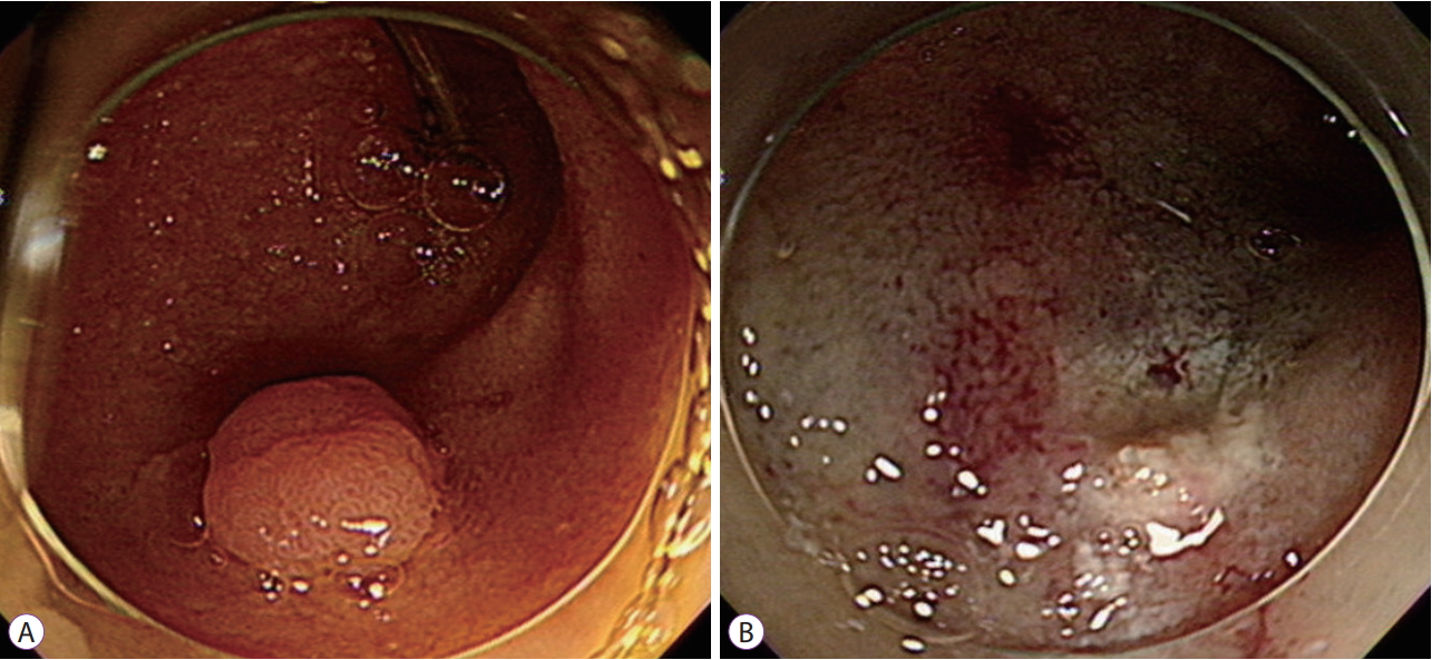
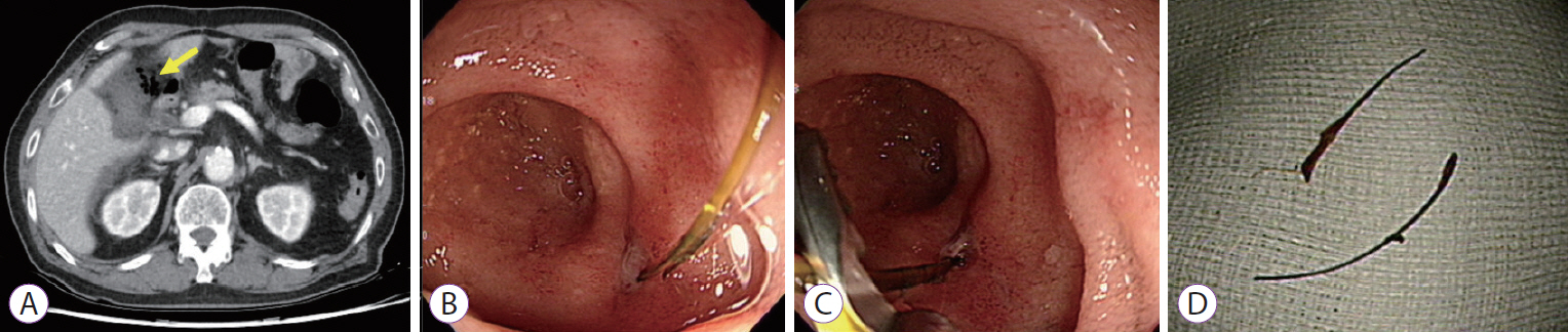
A 79-year-old man was referred to our hospital for removal of an adenoma in the afferent loop of a Billroth II anastomosis. Thirty-nine years earlier, he had undergone subtotal gastrectomy with Billroth II anastomosis because of gastric ulcer bleeding. His laboratory findings were unremarkable. Esophagogastroduodenoscopy revealed a subtly yellowish, elevated lesion 10 mm in diameter in the afferent loop of the Billroth II anastomosis. (Fig. 1A) The polyp was located in the distal portion approximately 3–4 cm apart from the anastomosis site. We resected the polyp by endoscopic mucosal resection (EMR), and the resection site was clear (Fig. 1B). No immediate adverse event related to the procedure was observed. After 2 days, however, the patient visited the emergency department because of severe epigastric pain. Abdominal computed tomography revealed fluid collection and free air around the afferent loop and right colon (Fig. 2A). Esophagogastroduodenoscopy revealed a sharp foreign body penetrating the EMR site (Fig. 2B). We meticulously examined the penetrating site using grasper forceps and found that the foreign body was directly penetrating the EMR-induced ulcer (Fig. 2C). We removed the foreign body using grasper forceps and closed the perforation site with endoclips (EZ Clip; Olympus, Tokyo, Japan), using single closure methods. The foreign body was a suture material used in the previous gastric surgery (Fig. 2D). The patient received conservative medical treatment without surgery. At 6-week follow-up, he had recovered completely without any symptoms.
Endoscopic resection is becoming the first choice of treatment for superficial duodenal epithelial neoplasia [1,2]. However, exposure to pancreatic juice or bile, thin duodenal wall, and retroperitoneal location increase the risk of perforation [3]. To prevent delayed perforation, complete closure of the mucosal defect after endoscopic resection is proposed, with favorable clinical outcomes [4]. We report a rare case in which a suture material used in the previous surgery caused delayed perforation of the EMR site. Complete closure of the mucosal defect after endoscopic resection is required to prevent delayed complications, even in the case of small duodenal polyps.
Fig. 1.
(A) An esophagogastroduodenoscopy image of a subtly yellowish, elevated lesion 10 mm in diameter in the afferent loop of a Billroth II anastomosis. (B) Clear resection site after endoscopic mucosal resection.
ce-2019-083f1.jpg
Fig. 2.
Images after 2 days. (A) A computed tomography scan showing fluid collection and free air around the afferent loop and right colon (yellow arrow). (B, C) An esophagogastroduodenoscopy image showing a sharp foreign body penetrating the endoscopic mucosal resection site. (D) The foreign body is a suture material used in the previous gastric surgery.
ce-2019-083f2.jpg
  • 1. Yahagi N, Kato M, Ochiai Y, et al. Outcomes of endoscopic resection for superficial duodenal epithelial neoplasia. Gastrointest Endosc 2018;88:676–682.ArticlePubMed
  • 2. Tomizawa Y, Ginsberg GG. Clinical outcome of EMR of sporadic, nonampullary, duodenal adenomas: a 10-year retrospective. Gastrointest Endosc 2018;87:1270–1278.ArticlePubMed
  • 3. Ochiai Y, Kato M, Kiguchi Y, et al. Current status and challenges of endoscopic treatments for duodenal tumors. Digestion 2019;99:21–26.ArticlePubMed
  • 4. Kato M, Ochiai Y, Fukuhara S, et al. Clinical impact of closure of the mucosal defect after duodenal endoscopic submucosal dissection. Gastrointest Endosc 2019;89:87–93.ArticlePubMed

Figure & Data

REFERENCES

    Citations

    Citations to this article as recorded by  

      • PubReader PubReader
      • ePub LinkePub Link
      • Cite
        CITE
        export Copy Download
        Close
        Download Citation
        Download a citation file in RIS format that can be imported by all major citation management software, including EndNote, ProCite, RefWorks, and Reference Manager.

        Format:
        • RIS — For EndNote, ProCite, RefWorks, and most other reference management software
        • BibTeX — For JabRef, BibDesk, and other BibTeX-specific software
        Include:
        • Citation for the content below
        Delayed Duodenal Perforation of an Endoscopic Mucosal Resection- Induced Ulcer due to a Foreign Body
        Clin Endosc. 2019;52(6):624-625.   Published online July 8, 2019
        Close
      • XML DownloadXML Download
      Figure
      • 0
      • 1
      Delayed Duodenal Perforation of an Endoscopic Mucosal Resection- Induced Ulcer due to a Foreign Body
      Image Image
      Fig. 1. (A) An esophagogastroduodenoscopy image of a subtly yellowish, elevated lesion 10 mm in diameter in the afferent loop of a Billroth II anastomosis. (B) Clear resection site after endoscopic mucosal resection.
      Fig. 2. Images after 2 days. (A) A computed tomography scan showing fluid collection and free air around the afferent loop and right colon (yellow arrow). (B, C) An esophagogastroduodenoscopy image showing a sharp foreign body penetrating the endoscopic mucosal resection site. (D) The foreign body is a suture material used in the previous gastric surgery.
      Delayed Duodenal Perforation of an Endoscopic Mucosal Resection- Induced Ulcer due to a Foreign Body

      Clin Endosc : Clinical Endoscopy Twitter Facebook
      Close layer
      TOP